

Titin based viscosity in ventricular physiology: an integrative investigation of PEVK-actin interactions
- PMID: 21708170
- PMCID: PMC3150406
- DOI: 10.1016/j.yjmcc.2011.06.006
Titin based viscosity in ventricular physiology: an integrative investigation of PEVK-actin interactions
Abstract
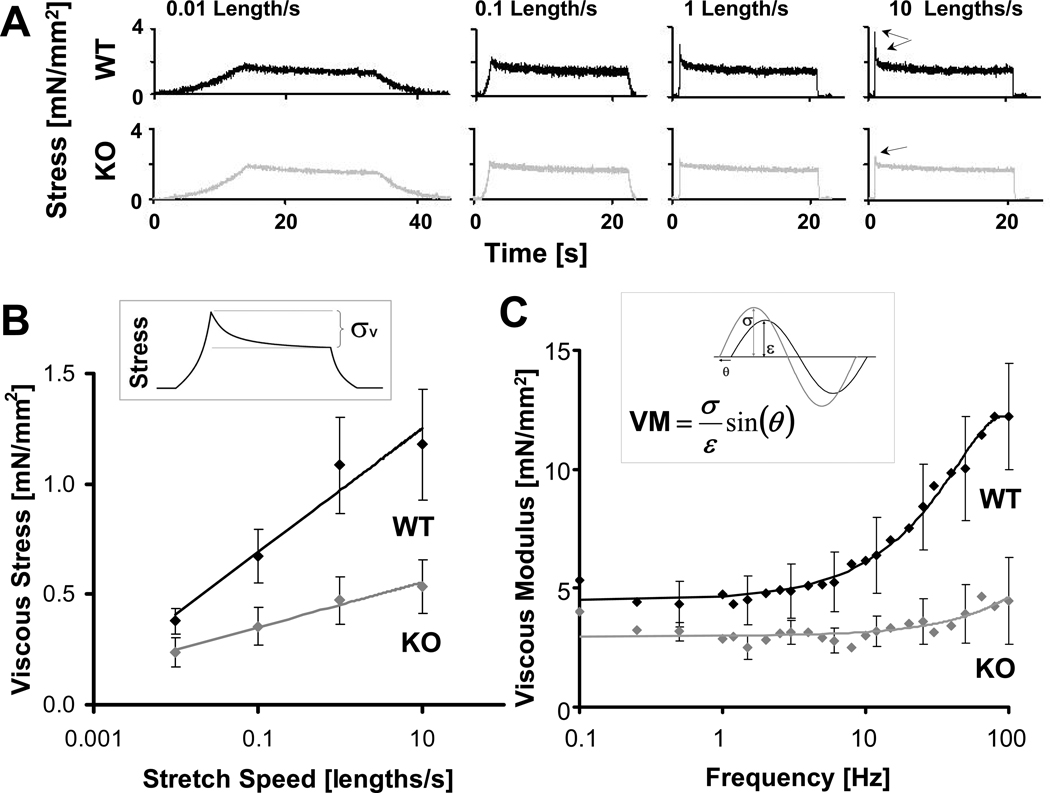
Viscosity is proposed to modulate diastolic function, but only limited understanding of the source(s) of viscosity exists. In vitro experiments have shown that the proline-glutamic acid-valine-lysine (PEVK) rich element of titin interacts with actin, causing a viscous force in the sarcomere. It is unknown whether this mechanism contributes to viscosity in vivo. We tested the hypothesis that PEVK-actin interaction causes cardiac viscosity and is important in vivo via an integrative physiological study on a unique PEVK knockout (KO) model. Both skinned cardiomyocytes and papillary muscle fibers were isolated from wildtype (WT) and PEVK KO mice and passive viscosity was examined using stretch-hold-release and sinusoidal analysis. Viscosity was reduced by ~60% in KO myocytes and ~50% in muscle fibers at room temperature. The PEVK-actin interaction was not modulated by temperature or diastolic calcium, but was increased by lattice compression. Stretch-hold and sinusoidal frequency protocols on intact isolated mouse hearts showed a smaller, 30-40% reduction in viscosity, possibly due to actomyosin interactions, and showed that microtubules did not contribute to viscosity. Transmitral Doppler echocardiography similarly revealed a 40% decrease in LV chamber viscosity in the PEVK KO in vivo. This integrative study is the first to quantify the influence of a specific molecular (PEVK-actin) viscosity in vivo and shows that PEVK-actin interactions are an important physiological source of viscosity.
Copyright © 2011 Elsevier Ltd. All rights reserved.
Figures


Similar articles
-
Interaction between PEVK-titin and actin filaments: origin of a viscous force component in cardiac myofibrils.Circ Res. 2001 Nov 9;89(10):874-81. doi: 10.1161/hh2201.099453. Circ Res. 2001. PMID: 11701614
-
Titin-actin interaction: PEVK-actin-based viscosity in a large animal.J Biomed Biotechnol. 2011;2011:310791. doi: 10.1155/2011/310791. Epub 2011 Nov 15. J Biomed Biotechnol. 2011. PMID: 22162634 Free PMC article.
-
Truncation of titin's elastic PEVK region leads to cardiomyopathy with diastolic dysfunction.Circ Res. 2009 Sep 11;105(6):557-64. doi: 10.1161/CIRCRESAHA.109.200964. Epub 2009 Aug 13. Circ Res. 2009. PMID: 19679835 Free PMC article.
-
Titin diversity--alternative splicing gone wild.J Biomed Biotechnol. 2010;2010:753675. doi: 10.1155/2010/753675. Epub 2010 Mar 21. J Biomed Biotechnol. 2010. PMID: 20339475 Free PMC article. Review.
-
Titin elasticity in the context of the sarcomere: force and extensibility measurements on single myofibrils.Adv Exp Med Biol. 2000;481:179-202; discussion 203-6. doi: 10.1007/978-1-4615-4267-4_11. Adv Exp Med Biol. 2000. PMID: 10987073 Review.
Cited by
-
The link between exercise and titin passive stiffness.Exp Physiol. 2017 Sep 1;102(9):1055-1066. doi: 10.1113/EP086275. Epub 2017 Jul 31. Exp Physiol. 2017. PMID: 28762234 Free PMC article. Review.
-
Impact of lengthening velocity on the generation of eccentric force by slow-twitch muscle fibers in long stretches.Pflugers Arch. 2024 Oct;476(10):1517-1527. doi: 10.1007/s00424-024-02991-4. Epub 2024 Jul 24. Pflugers Arch. 2024. PMID: 39043889 Free PMC article.
-
Cell- and molecular-level mechanisms contributing to diastolic dysfunction in HFpEF.J Appl Physiol (1985). 2015 Nov 15;119(10):1228-32. doi: 10.1152/japplphysiol.01168.2014. Epub 2015 Apr 24. J Appl Physiol (1985). 2015. PMID: 25911687 Free PMC article. Review.
-
The Effects of Mechanical Preload on Transmural Differences in Mechano-Calcium-Electric Feedback in Single Cardiomyocytes: Experiments and Mathematical Models.Front Physiol. 2020 Mar 17;11:171. doi: 10.3389/fphys.2020.00171. eCollection 2020. Front Physiol. 2020. PMID: 32256377 Free PMC article.
-
hsa-miR-548v controls the viscoelastic properties of human cardiomyocytes and improves their relaxation rates.JCI Insight. 2024 Jan 2;9(3):e161356. doi: 10.1172/jci.insight.161356. JCI Insight. 2024. PMID: 38165745 Free PMC article.
References
-
- Chung CS, Kovács SJ. Physical determinants of left ventricular isovolumic pressure decline: model prediction with in vivo validation. Am J Physiol Heart Circ Physiol. 2008 Apr 1;294(4):H1589–H1596. - PubMed
-
- Kass DA, Bronzwaer JGF, Paulus WJ. What mechanisms underlie diastolic dysfunction in heart failure? Circ Res. 2004 Jun 25;94(12):1533–1542. - PubMed
-
- Tskhovrebova L, Trinick J, Sleep JA, Simmons RM. Elasticity and unfolding of single molecules of the giant muscle protein titin. Nature. 1997 May 15;387(6630):308–312. - PubMed
Publication types
MeSH terms
Substances
Grants and funding
LinkOut - more resources
Full Text Sources
Other Literature Sources
Research Materials
