. 2011 Aug 26;411(4):744-64.
doi: 10.1016/j.jmb.2011.06.021. Epub 2011 Jun 25.

Intrasubunit and intersubunit interactions controlling assembly of active synaptic complexes during Hin-catalyzed DNA recombination

Affiliations

Intrasubunit and intersubunit interactions controlling assembly of active synaptic complexes during Hin-catalyzed DNA recombination

John K Heiss et al. J Mol Biol. .

Abstract

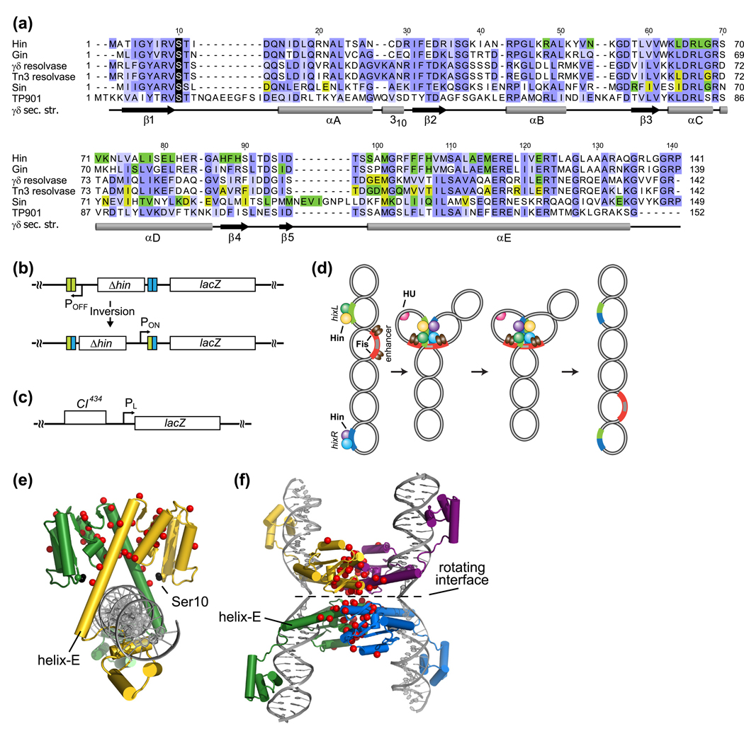
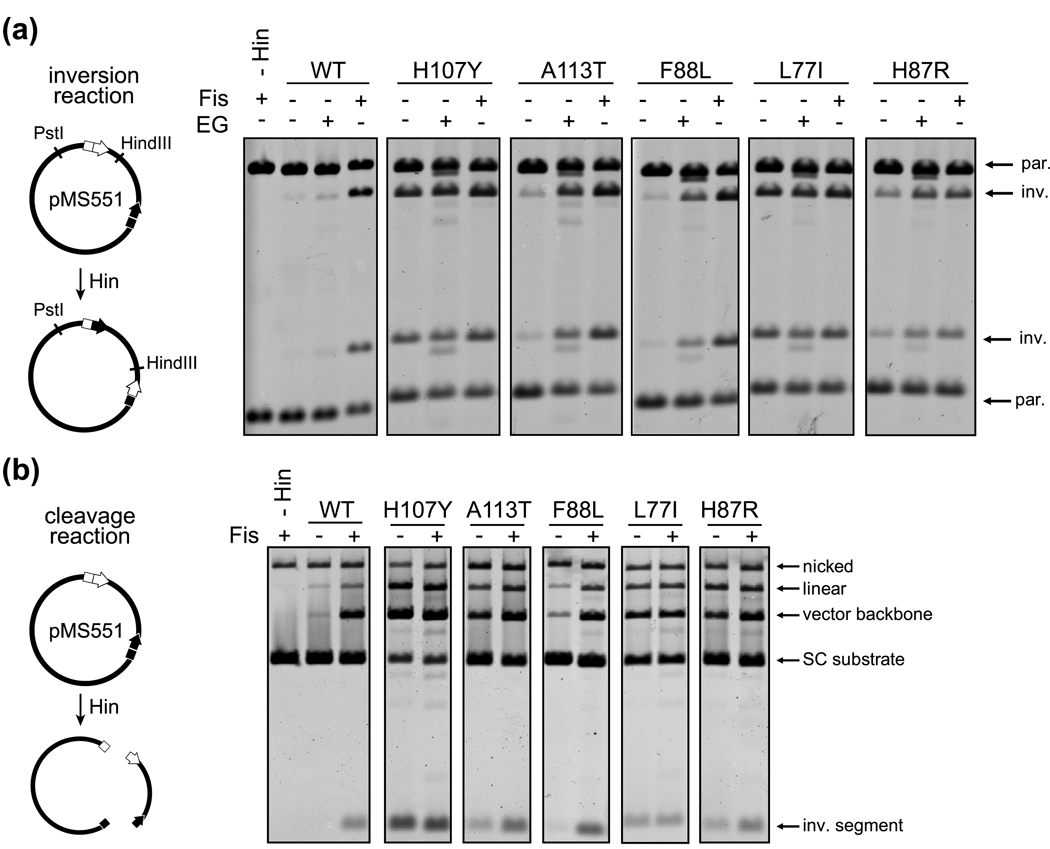
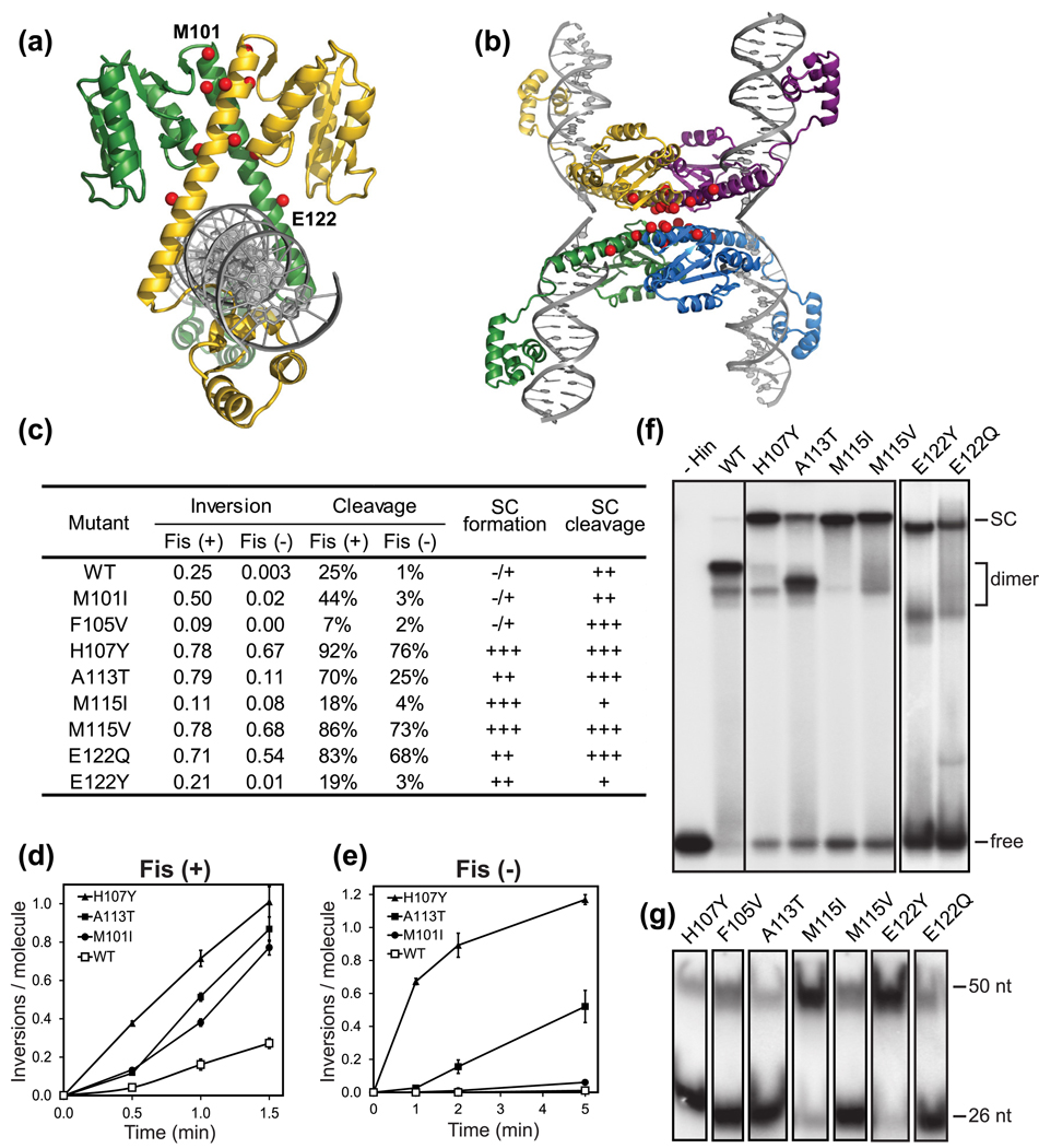
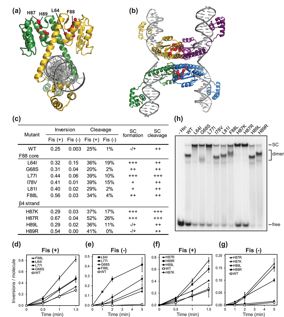
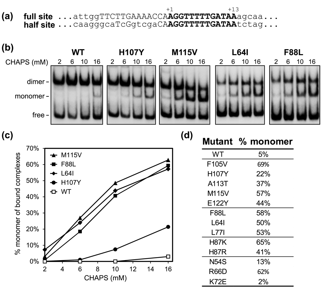
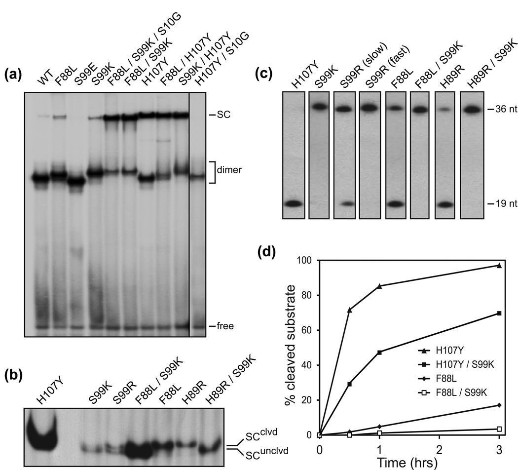
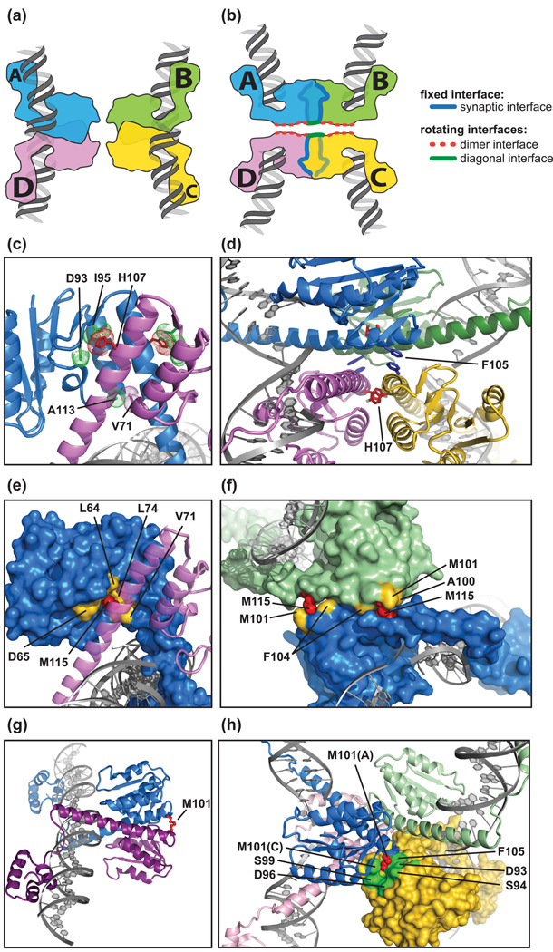
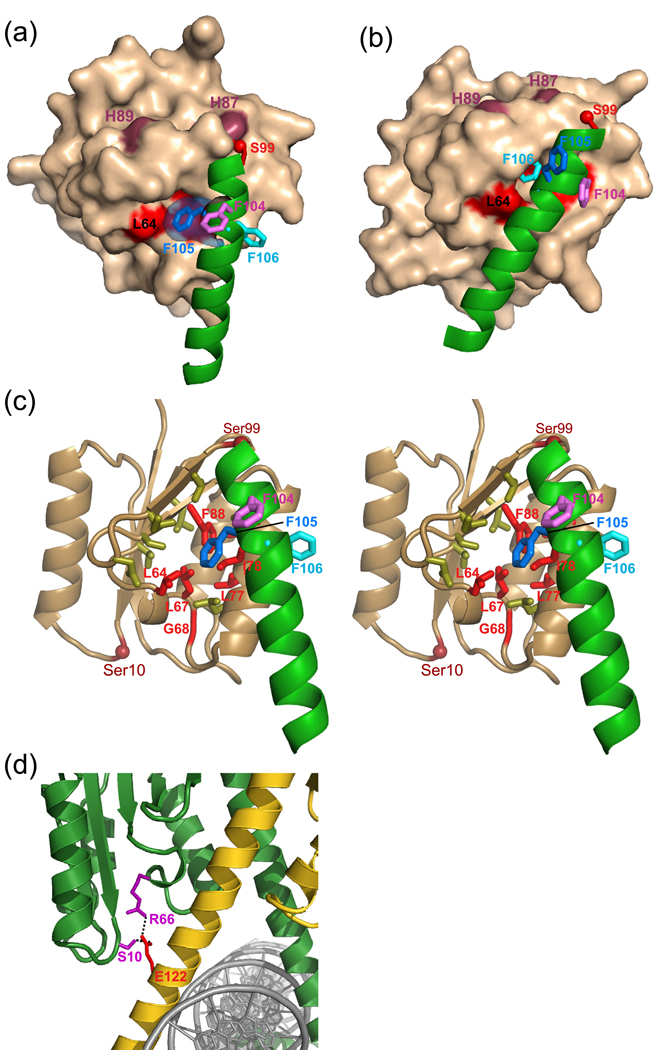
Serine recombinases, which generate double-strand breaks in DNA, must be carefully regulated to ensure that chemically active DNA complexes are assembled correctly. In the Hin-catalyzed site-specific DNA inversion reaction, two inversely oriented recombination sites on the same DNA molecule assemble into a synaptic complex that uniquely generates inversion products. The Fis-bound recombinational enhancer, together with topological constraints directed by DNA supercoiling, functions to regulate Hin synaptic complex formation and activity. We have isolated a collection of gain-of-function mutants in 22 positions within the catalytic and oligomerization domains of Hin using two genetic screens and by site-directed mutagenesis. One genetic screen measured recombination in the absence of Fis and the other assessed SOS induction as a readout of increased DNA cleavage. These mutations, together with molecular modeling, identify important sites of dynamic intrasubunit and intersubunit interactions that regulate assembly of the active tetrameric recombination complex. Of particular interest are interactions between the oligomerization helix (helix E) and the catalytic domain of the same subunit that function to hold the dimer in an inactive state in the absence of the Fis/enhancer system. Among these is a relay involving a triad of phenylalanines that are proposed to switch positions during the transition from dimers to the catalytically active tetramer. Novel Hin mutants that generate synaptic complexes that are blocked at steps prior to DNA cleavage are also described.


Figures

Fig. 1
Fig. 1
Serine recombinases and the Hin reaction. (a) Sequence of the conserved catalytic domain and helix E region of Hin and other serine recombinases. Gin is a related DNA invertase, γδ, Tn3, and Sin resolvases catalyze deletion, and the TP901 integrase catalyzes phage integration/excision. Common and similar residues are highlighted in blue and light blue shading, respectively, and the active site serines are demarked with black shading. Residues with green shading highlight locations where single substitutions have been reported to confer hyperactivity in the listed recombinases. Yellow shading on resolvases and Sin signify residues where hyperactivity is observed only in the presence of a second mutation. For resolvases and Sin, hyperactivity typically reflects recombination of substrates lacking accessory sites on one of the two recombination sites. Crystal structures of dimeric and tetrameric γδ resolvase, and Sin (regulatory tetramer) and TP901 (tetramer) are available; the designated secondary structure is from the γδ resolvase tetramer (1ZR4.pdb), which fits well with secondary structure predictions and modeling of Hin. (b) Flagellin controlling locus in Salmonella enterica serovar Typhimurium. A 996 bp invertible DNA segment contains the hin gene and an outward facing Eσ promoter, which transcribes the fljB (H2) flagellin gene and a repressor of an unlinked flagellin gene in one orientation. The drawing schematically depicts the λfla-lac406 reporter construct used to monitor Hin-catalyzed inversion in vivo where the hin gene is inactivated, and lacZ is substituted in place of the repressor. hix recombination sites are denoted in green and blue. (c) Schematic representation of the reporter used for monitoring SOS induction by Hin. (d) Schematic representation of the Hin recombination pathway on a plasmid substrate. Hin binds to the hix sites and Fis dimers bind to the recombinational enhancer. The HU bending protein facilitates assembly of the invertasome in which DNA cleavage, subunit rotation, and ligation occur to invert the intervening segment of DNA (adapted from ref.). (e and f) Structural models of the Hin dimer based on γδ resolvase (1GDT) and the rotationally competent, DNA cleaved, Hin synaptic tetramer based on γδ resolvase (2GM4). The locations of regulatory mutants discussed in this report are designated with red spheres and the catalytic serine 10 with a black sphere.
Fig. 2
Fig. 2
In vitro DNA inversion and cleavage reactions on supercoiled plasmids by wild type and representative hyperactive Hin mutants. (a) In vitro DNA inversion reactions. Fis and 15% ethylene glycol (EG) were added as designated. After 2 min Hin reactions, plasmids were digested with Pst I and HindIII to distinguish the orientation of the invertible segment, as illustrated in the drawing, and electrophoresed in an agarose gel. (par) designate products representing the parental orientation; (inv) designate products representing the inverted orientation. (b) Hin-catalyzed DNA cleavage reactions. The products of Hin cleavage reactions (2 min) were treated with proteinase K and SDS and electrophoresed in an agarose gel. The locations of the supercoiled (SC) substrate, nicked plasmid, linear plasmid containing a single double-strand break, and the vector backbone plus invertible (inv) segment resulting from two double-strand breaks are designated.
Fig. 3
Fig. 3
Hyperactivating Hin mutations in helix-E. (a) Location of the helix-E mutations (red spheres) on the Hin dimer and (b) DNA-cleaved tetramer models. (c) Summary of in vitro activities by helix-E hyperactive mutants. Inversion rates with and without Fis are given as inversions/plasmid molecule/min, and cleavage rates with and without Fis were calculated from the number of double and single cleaved plasmids generated per minute relative to the total DNA, excluding the open circular form. Synaptic complex (SC) formation was determined from the amount of synaptic complexes relative to the total Hin-bound DNA (panel (f)). (+++) indicates >75% of the bound DNA is assembled into synaptic complexes, (++) indicates 30–75%, (+) indicates 10–30%, and (−/+) indicates ≤5 %. SC cleavage refers to hix site cleavage as determined by extracting synaptic complexes in the presence of SDS and quantifying the amount of cleaved DNA by denaturing PAGE (panel (g)). (+++) indicates >75% of the hix sites were cleaved, (++) indicates 25–75% cleaved, (+) indicates 5–25% cleaved, −/+ indicates <5% cleaved. All numbers are mean values from at least 3 experiments. (d and e) Representative in vitro DNA inversion reactions in the presence or absence of Fis. (f) Representative synaptic complex assays formed with 36 bp hixL sites. Variable migrations of the dimer-bound hix complexes are commonly observed and appear to reflect electrophoretic differences caused by the amino acid substitutions and perhaps from conformational differences. The slightly faster migrations of the tetrameric synaptic complexes (SC) formed by Hin-M115I and Hin-E122Q relative to the other mutants are resolved better upon longer electrophoresis (e.g., Fig. 6b). (g) DNA cleavage by Hin mutants within gel-isolated synaptic complexes. Synaptic complexes from native polyacrylamide gels as in panel (f) were extracted in the presence of SDS and subjected to denaturing PAGE. Uncleaved (50 nt) and cleaved (26 nt) 5’-32P-labeled products are denoted.
Fig. 4
Fig. 4
Hyperactivating Hin mutations in the Phe88 hydrophobic core and β-4 strand region. (a) Location of the Phe88 core and β-4 mutations (red spheres) on the Hin dimer and (b) DNA-cleaved tetramer models. (c) Summary of in vitro activities by the mutants. Activities are designated as in Fig. 3c. (d–g) Representative in vitro DNA inversion reactions in the presence or absence of Fis. (h) Native polyacrylamide gel of synaptic complex reactions.
Fig. 5
Fig. 5
Stability of Hin mutant dimers to CHAPS as evaluated by hixL half-site binding. (a) Sequence of the hixL half-site substrate used in the DNA binding assays. (b) Gel mobility shift assays on the 183 bp 32P-labeled hix half site fragments with a constant amount of Hin-wt and representative mutants and different concentration of CHAPS in the binding buffer. The locations of the dimer and monomer complexes along with the unbound substrate (free) are denoted. (c) The % monomer complex relative to the bound (monomer + dimer) complexes is plotted for the different CHAPS concentrations for the wild type and mutants shown in panel b. (d) Summary of the % monomer bound at 16 mM CHAPS for different Hin mutants. Values represent the mean of at least 3 experiments.
Fig. 6
Fig. 6
Properties of synaptic complexes by Hin mutants containing Ser99 mutations. (a) Native PAGE of synaptic complex reactions by Hin mutants. S10G inactivates the active-site serine. (b) Electrophoretic migration differences of DNA cleaved and uncleaved synaptic complexes (SC) formed on 36 bp hixL fragments. (c) Synaptic complexes were extracted in SDS buffer and subjected to denaturing PAGE. S99R (slow) and (fast) refer to the electrophoretic migration of the isolated synaptic complexes as in panel (b). (d) Rates of DNA cleavage on 36 bp hixL fragments by Hin-H107Y and F88L mutants with and without S99K. Reactions were quenched with SDS at various times, subjected to denaturing PAGE, and the amounts of cleaved and uncleaved DNAs quantified. Note that rates of formation of synaptic complexes by H107Y and F88L/S99K, as assayed by native PAGE, are similar and reach a maximum yield within several minutes.
Fig. 7
Fig. 7
Structural interpretations of helix-E hyperactivating mutations. Schematic representations of the (a) Hin dimer and (b) DNA-cleaved tetramer. Note the chain designations (A–D), coloring of subunits, and the different subunit interfaces in the tetramer. (c) Hin dimer model highlighting the side chains of His107 along with Ala113 and Val71. (d) DNA-cleaved Hin tetramer model highlighting the stacking of residue 107 side chains (shown as red tyrosines as in H107Y) from subunits across the new synaptic interface and the dark blue Phe105 side chains across the rotating interface between diagonally-oriented subunits. See Supplementary Movie 1. (e) Hin dimer model highlighting the connection between Met115 (red spheres) on subunit D and a set of residues (yellow) across the dimer interface on the surface of the subunit A. (f) DNA-cleaved tetramer model highlighting the connections between Met115 (red) on subunit A and residues (gold) on the synapsed subunit B. The rotating subunit C and D pair have been removed for clarity, revealing the rotating interface on the plane of the page. See Supplementary Movie 2. (g) Hin dimer model showing locations of Met101 at the top of the dimer interface. (i) Open Hin tetramer intermediate model showing Met101 (red) from subunit A engaged in a set of connections with residues from the diagonally-opposed subunit C. In the final DNA-cleaved tetramer complex, Met101 is primarily engaged in contacts with the newly synapsed subunit near the edge of the rotating interface to help stabilize the rotating subunit pair but also comprises part of the rotating interface (panel f).
Fig. 8
Fig. 8
The helix-E phenylalanine triad and Phe88 core hyperactivating mutations. (a) Subunit A of the Hin dimer model depicting a surface representation of the catalytic domain (residues 1–98), Ser99, and helix E (residues 100–120) as a green ribbon. The side chains of phenylalanines 104, 105 (with transparent surface inserted within the Phe88 pocket of the catalytic domain), and 106 are shown along with selected residues in the catalytic domain. The red surfaces of Phe88 and Ile78 are visible behind Phe105. (b) Subunit A of the DNA-cleaved tetramer model in a similar view as in panel a. Note that Phe105 is rotated out of the Phe88 pocket and Phe106 is now inserted. See Supplementary Movie 3. (c) Stereo pair of the Hin dimer model looking into the Phe88 core showing locations of mostly hydrophobic side chains. Residues where hyperactivating mutations were isolated are denoted in red and labeled. The positions of side chains should be considered only as approximate. (d) Potential interactions between the side chains of Gln122 on subunit C and Arg66 and the active site residue Ser10 on subunit B on the Hin dimer model.

Similar articles

Cited by

References

    1. Grindley NDF, Whiteson KL, Rice PA. Mechanisms of site-specific recombination. Annu. Rev. Biochem. 2006;75:567–605. - PubMed
    1. Johnson RC. Bacterial Site-specific DNA inversion systems. In: Craig NL, Craigie R, Gellert M, Lambowitz AM, editors. Mobile DNA II. Washington, D.C.: ASM Press; 2002. pp. 230–271.
    1. Zieg J, Silverman M, Hilmen M, Simon M. Recombinational switch for gene expression. Science. 1977;196:170–172. - PubMed
    1. Yang W, Steitz TA. Crystal structure of the site-specific recombinase gamma delta resolvase complexed with a 34 bp cleavage site. Cell. 1995;82:193–207. - PubMed
    1. Johnson RC, Simon MI. Hin-mediated site-specific recombination requires two 26 bp recombination sites and a 60 bp recombinational enhancer. Cell. 1985;41:781–791. - PubMed

Publication types

MeSH terms

LinkOut - more resources