

Cholinergic modulation of cognition: insights from human pharmacological functional neuroimaging
- PMID: 21708219
- PMCID: PMC3382716
- DOI: 10.1016/j.pneurobio.2011.06.002
Cholinergic modulation of cognition: insights from human pharmacological functional neuroimaging
Abstract
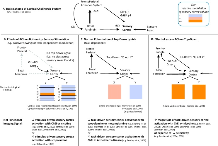
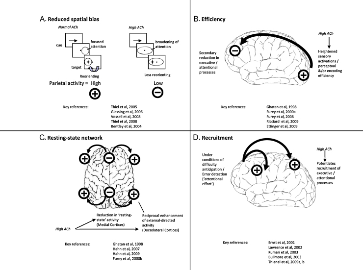
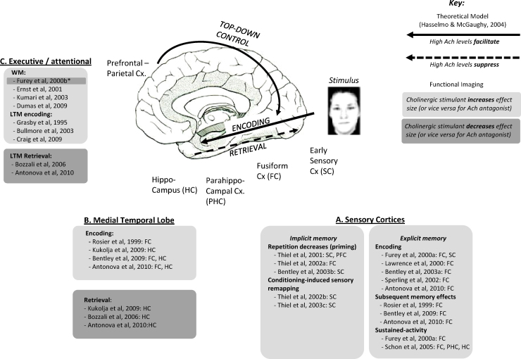
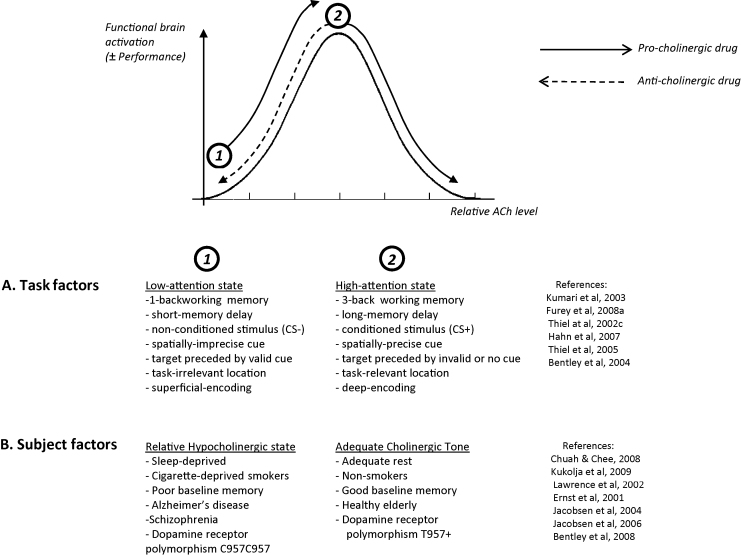
Evidence from lesion and cortical-slice studies implicate the neocortical cholinergic system in the modulation of sensory, attentional and memory processing. In this review we consider findings from sixty-three healthy human cholinergic functional neuroimaging studies that probe interactions of cholinergic drugs with brain activation profiles, and relate these to contemporary neurobiological models. Consistent patterns that emerge are: (1) the direction of cholinergic modulation of sensory cortex activations depends upon top-down influences; (2) cholinergic hyperstimulation reduces top-down selective modulation of sensory cortices; (3) cholinergic hyperstimulation interacts with task-specific frontoparietal activations according to one of several patterns, including: suppression of parietal-mediated reorienting; decreasing 'effort'-associated activations in prefrontal regions; and deactivation of a 'resting-state network' in medial cortex, with reciprocal recruitment of dorsolateral frontoparietal regions during performance-challenging conditions; (4) encoding-related activations in both neocortical and hippocampal regions are disrupted by cholinergic blockade, or enhanced with cholinergic stimulation, while the opposite profile is observed during retrieval; (5) many examples exist of an 'inverted-U shaped' pattern of cholinergic influences by which the direction of functional neural activation (and performance) depends upon both task (e.g. relative difficulty) and subject (e.g. age) factors. Overall, human cholinergic functional neuroimaging studies both corroborate and extend physiological accounts of cholinergic function arising from other experimental contexts, while providing mechanistic insights into cholinergic-acting drugs and their potential clinical applications.
Copyright © 2011 Elsevier Ltd. All rights reserved.
Figures
Similar articles
-
Unraveling the attentional functions of cortical cholinergic inputs: interactions between signal-driven and cognitive modulation of signal detection.Brain Res Brain Res Rev. 2005 Feb;48(1):98-111. doi: 10.1016/j.brainresrev.2004.08.006. Brain Res Brain Res Rev. 2005. PMID: 15708630 Review.
-
More attention must be paid: the neurobiology of attentional effort.Brain Res Rev. 2006 Aug;51(2):145-60. doi: 10.1016/j.brainresrev.2005.11.002. Epub 2006 Mar 13. Brain Res Rev. 2006. PMID: 16530842 Review.
-
Feature-Selective Attentional Modulations in Human Frontoparietal Cortex.J Neurosci. 2016 Aug 3;36(31):8188-99. doi: 10.1523/JNEUROSCI.3935-15.2016. J Neurosci. 2016. PMID: 27488638 Free PMC article.
-
Neural correlates of successful emotional episodic encoding and retrieval: An SDM meta-analysis of neuroimaging studies.Neuropsychologia. 2020 Jun;143:107495. doi: 10.1016/j.neuropsychologia.2020.107495. Epub 2020 May 13. Neuropsychologia. 2020. PMID: 32416099
-
Acetylcholine and attention.Behav Brain Res. 2011 Aug 10;221(2):430-42. doi: 10.1016/j.bbr.2010.11.033. Epub 2010 Nov 23. Behav Brain Res. 2011. PMID: 21108972 Review.
Cited by
-
Long-term memory search across the visual brain.Neural Plast. 2012;2012:392695. doi: 10.1155/2012/392695. Epub 2012 Jul 19. Neural Plast. 2012. PMID: 22900206 Free PMC article. Review.
-
Infralimbic cortical glutamate output is necessary for the neural and behavioral consequences of chronic stress.Neurobiol Stress. 2020 Nov 23;13:100274. doi: 10.1016/j.ynstr.2020.100274. eCollection 2020 Nov. Neurobiol Stress. 2020. PMID: 33344727 Free PMC article.
-
Cholinergic enhancement reduces functional connectivity and BOLD variability in visual extrastriate cortex during selective attention.Neuropharmacology. 2013 Jan;64(1):305-13. doi: 10.1016/j.neuropharm.2012.07.003. Epub 2012 Aug 18. Neuropharmacology. 2013. PMID: 22906685 Free PMC article. Clinical Trial.
-
Pharmacological targeting of cognitive impairment in depression: recent developments and challenges in human clinical research.Transl Psychiatry. 2022 Nov 17;12(1):484. doi: 10.1038/s41398-022-02249-6. Transl Psychiatry. 2022. PMID: 36396622 Free PMC article. Review.
-
Pharmacological dissociation of novelty responses in the human brain.Cereb Cortex. 2014 May;24(5):1351-60. doi: 10.1093/cercor/bhs420. Epub 2013 Jan 10. Cereb Cortex. 2014. PMID: 23307638 Free PMC article.
References
-
- Adachi T., Inanami O., Ohno K., Sato A. Responses of regional cerebral blood flow following focal electrical stimulation of the nucleus basalis of Meynert and the medial septum using the [14C]iodoantipyrine method in rats. Neurosci. Lett. 1990;112(2–3):263–268. - PubMed
-
- Anagnostaras S.G., Murphy G.G., Hamilton S.E., Mitchell S.L., Rahnama N.P., Nathanson N.M., Silva A.J. Selective cognitive dysfunction in acetylcholine M1 muscarinic receptor mutant mice. Nat. Neurosci. 2003;6(1):51–58. - PubMed
-
- Antonova E., Parslow D., Brammer M., Simmons A., Williams S., Dawson G.R., Morris R.G. Scopolamine disrupts hippocampal activity during allocentric spatial memory in humans: an fMRI study using a virtual reality analogue of the Morris Water Maze. J. Psychopharmacol. 2010 September 7 (Epub ahead of print) - PubMed
-
- Aoyagi M., Meyer J.S., Deshmukh V.D., Ott E.O., Tagashira Y., Kawamura Y., Matsuda M., Achari A.N., Chee A.N. Central cholinergic control of cerebral blood flow in the baboon. Effect of cholinesterase inhibition with neostigmine on autoregulation and CO2 responsiveness. J. Neurosurg. 1975;43(6):689–705. - PubMed
Publication types
MeSH terms
Substances
Grants and funding
LinkOut - more resources
Full Text Sources
Medical
