Review
. 2011 Sep 1;94(4):307-46.
doi: 10.1016/j.pneurobio.2011.06.001. Epub 2011 Jun 17.

Beyond faithful conduction: short-term dynamics, neuromodulation, and long-term regulation of spike propagation in the axon

Affiliations
Review

Beyond faithful conduction: short-term dynamics, neuromodulation, and long-term regulation of spike propagation in the axon

Dirk Bucher et al. Prog Neurobiol. .

Abstract

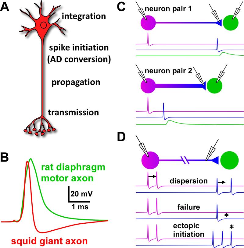
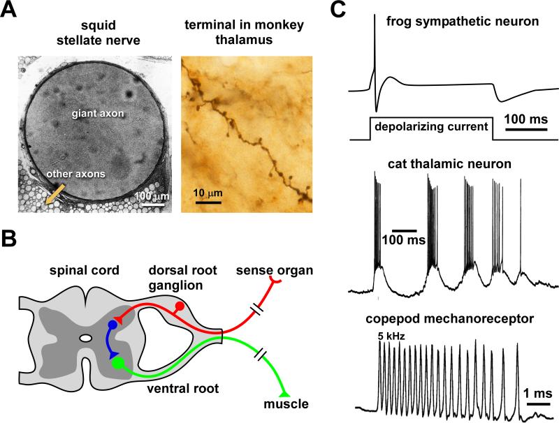
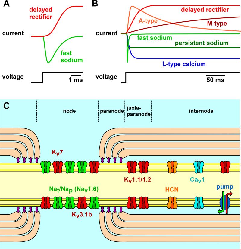
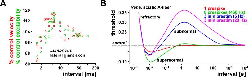
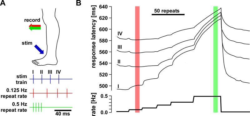
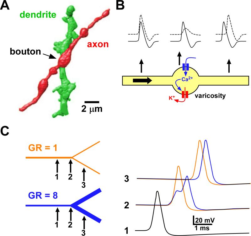
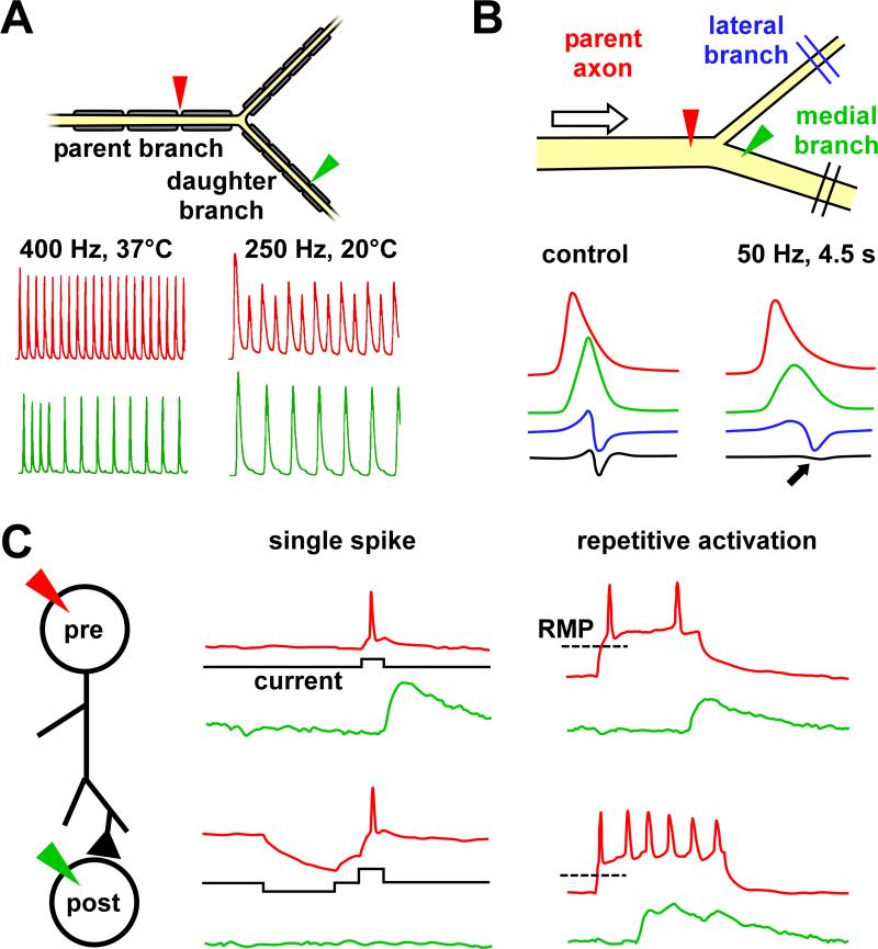
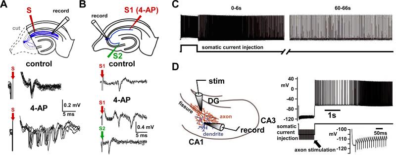
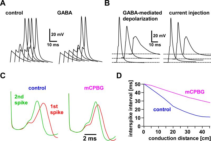
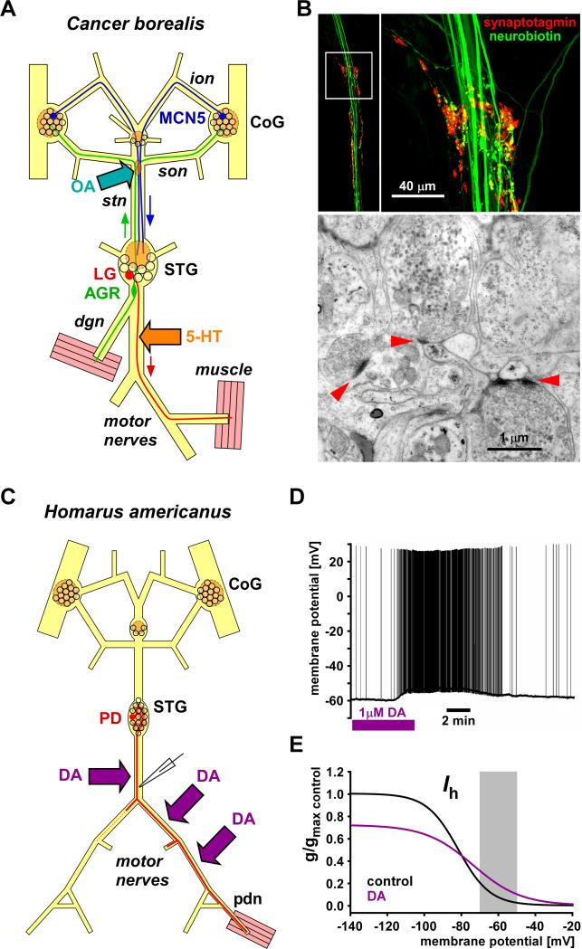
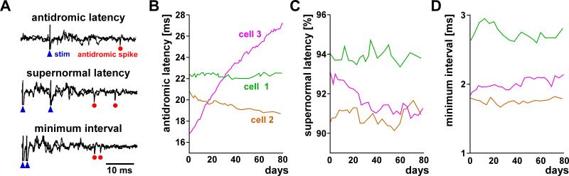
Most spiking neurons are divided into functional compartments: a dendritic input region, a soma, a site of action potential initiation, an axon trunk and its collaterals for propagation of action potentials, and distal arborizations and terminals carrying the output synapses. The axon trunk and lower order branches are probably the most neglected and are often assumed to do nothing more than faithfully conducting action potentials. Nevertheless, there are numerous reports of complex membrane properties in non-synaptic axonal regions, owing to the presence of a multitude of different ion channels. Many different types of sodium and potassium channels have been described in axons, as well as calcium transients and hyperpolarization-activated inward currents. The complex time- and voltage-dependence resulting from the properties of ion channels can lead to activity-dependent changes in spike shape and resting potential, affecting the temporal fidelity of spike conduction. Neural coding can be altered by activity-dependent changes in conduction velocity, spike failures, and ectopic spike initiation. This is true under normal physiological conditions, and relevant for a number of neuropathies that lead to abnormal excitability. In addition, a growing number of studies show that the axon trunk can express receptors to glutamate, GABA, acetylcholine or biogenic amines, changing the relative contribution of some channels to axonal excitability and therefore rendering the contribution of this compartment to neural coding conditional on the presence of neuromodulators. Long-term regulatory processes, both during development and in the context of activity-dependent plasticity may also affect axonal properties to an underappreciated extent.


Figures

Fig. 1
Fig. 1
Action potential conduction in axons of spiking neurons. A: Schematic representation of a neuron with proximal integration of synaptic input and spike initiation. Spikes are propagated along the axon into distal terminals where depolarization results in transmitter release. B: Typical spike waveforms obtained from intracellular recordings. C: Schematic of conduction delay. The propagation time introduces a latency between spike initiation and postsynaptic responses potentially much larger than the synaptic delay, and very different across different pairs of neurons. D: Changes of temporal patterns between proximal and distal recording sites introduced by the process of propagation, including temporal dispersion, spike failures, and ectopic spike initiation. B is modified from Hodgkin and Huxley, 1939, and David et al., 1995.
Fig. 2
Fig. 2
Diversity of axon morphology and neuronal firing patterns. A: Different axon diameters. The left panel shows a cross section of a squid (Sepioteuthis lessoniana) stellar nerve with vastly different axon sizes. The right panel shows a small-bouton spinous axon terminal of a cortico-thalamic neuron from a macaque monkey, filled with a retrograde tracer. B: Schematic of spinal and dorsal root ganglion neurons. Local interneurons (blue) have fairly short axons, whereas the axons of sensory (red) and motor (green) neurons can be > 1 m long. C: Different firing behavior. The upper panel shows a fast-adapting sympathetic neuron. These neurons often only fire single spikes in response to sustained depolarization, due to substantial M-type potassium currents and slow after-hyperpolarization. The middle panel shows a thalamic neuron during spindle oscillation. The lower panel shows the extreme high-frequency response of a copepod antennal mechanoreceptor to a water jet. A is modified from Lee et al., 1994, and Miyashita et al., 2007; C is modified from Jones, 1985, Contreras and Steriade, 1996, and Fields and Weissburg, 2004.
Fig. 3
Fig. 3
Diversity of ionic currents in axons. A: Fast sodium and delayed rectifier potassium currents in the squid giant axon, as elicited in response to a depolarizing voltage step. B: Cartoon of current responses to depolarizing voltage steps in an axon with a more complex complement of channels which activate and inactivate with very different time constants. C: A range of different ion channels found in either central or peripheral myelinated axons, distributed differentially between node, juxtaparanode, and internode. A is modified from Hodgkin and Huxley, 1952.
Fig. 4
Fig. 4
Activity-dependent changes in axonal excitability and conduction delay in earthworm and frog axons. A: Concomitant relative changes of conduction velocity and excitability as a function of interval between paired pulses in an earthworm axon. Excitability was measured as the threshold for spike initiation at the stimulus site. At small intervals, conduction velocity and excitability was reduced. At larger intervals, velocity and excitability was increased and returned to control values only at intervals greater than 100 ms. B: Triphasic changes in excitability and their dependence on conditioning regime in frog sciatic axons. With a single conditioning spike (red), the threshold for the initiation of a second spike was increased at small intervals (relative refractory period), and decreased at larger intervals (supernormal period). When the axon was conditioned with 8 impulses at 450 Hz (green), the supernormal period was increased and followed by a period of slightly increased threshold (subnormal). The subnormal period was dramatically increased when the axon was conditioned with 5 Hz (blue) or 20 Hz (magenta) stimulation sustained over 3 minutes. A is modified from Bullock, 1951; B is modified from Raymond, 1979.
Fig. 5
Fig. 5
Afterpotentials and activity-dependent changes in axonal excitability. A: Spike recorded from a myelinated motor axon innervating the diaphragm in the rat. B: Enlarged view of the voltage range around resting membrane potential and expanded time base of the same trace shown in A. A depolarizing afterpotential (DAP) is followed by an after-hyperpolarization (AHP). C: The relationship between afterpotentials and excitability changes in a computational model of a myelinated axon, calculated for three different axon diameters. Note that the changes associated with the relative refractory period at short intervals (~2-4 ms) are not reflected in the membrane potential. However, later changes in threshold follow the DAP and AHP quite well. The difference in DAP between axons of different sizes was due to the fact that passive capacitive charging depends on diameter. A and B are modified from David et al., 1995; C is modified from McIntyre et al., 2002.
Fig. 6
Fig. 6
Changes in spike shape during repetitive activity and changes in baseline membrane potential A: In a Hodgkin-Huxley type axon model with only fast sodium and delayed rectifier potassium currents, sodium channel inactivation leads to a reduction in spike amplitude (red arrows). B: Conduction velocity in the same model is well correlated with the peak voltage of spikes, in that spikes with reduced amplitude are slower. C: In hippocampal mossy fibers, inactivation of A-type potassium channels leads to activity-dependent spike broadening. The overlaid traces shown are from a 50 Hz train. D: The magnitude of the increase in spike duration in mossy fibers is dependent on stimulation frequency. E: In a lobster stomatogastric axon, depolarizing and hyperpolarizing current injections during ongoing burst activity change spike amplitudes, spike durations, and the dynamics of both. Shown are overlaid spikes from single bursts under each condition. Depolarizing current injection (left) decreases initial spike amplitude (red bar) and increases the frequency dependent change in amplitude (green bar), compared to control (middle panel). Initial spike duration (blue bar) is prolonged compared to control, but increases very little over the course of the burst (magenta bar). Hyperpolarizing current injection (right) increases spike amplitude and decreases frequency-dependent reduction. Initial spike duration is reduced compared to control but increases substantially over the course of the burst. A and B are modified from Moradmand and Goldfinger, 1995. C and D are modified from Geiger and Jonas, 2000. E is modified from Ballo and Bucher, 2009.
Fig. 7
Fig. 7
Spatial variation and entrainment of intervals between spikes propagated at different velocities. A: Conduction velocity as a function of stimulus interval in a Hodgkin-Huxley axon model stimulated with paired pulses. Velocities were calculated from measurements at a distance of 9 length constants from the stimulation site. The conditioning spike traveled at a velocity of 6.54 m/s (dashed red line). The second spike was slowed when elicited during the relative refractory period. With increasing intervals, conduction became first supernormal and then subnormal (green line). B: When the interstimulus interval was varied between 5 and 23 ms in the same model axon, the interspike interval changed as a function of distance from the stimulation site. Over distance, spikes elicited during the relative refractory period and in the early supernormal period converge on an interval that marks the transition between refractory and supernormal periods, where the conduction velocities of conditioning and second spike are identical (blue ellipse). C: Impulse entrainment in a rabbit efferent visual cortex neuron. Thalamic antidromic stimulation at a distance of 14.9 mm was used to evoke spikes in cortical extracellular recordings. Recorded spike intervals were a constant 1.7 ms, despite the fact that stimulus intervals were varied between 0.9 and 2.0 ms (10 pairs of stimuli as shown in the lower scheme). A and B are modified from Moradmand and Goldfinger, 1995; C is modified from Kocsis et al.,1979.
Fig. 8
Fig. 8
Membrane potential, spike shape, and conduction delay in a crustacean stomatogastric axon. A: Single burst recorded from the pyloric dilator (PD) neuron axon in the motor nerves during ongoing rhythmic pyloric activity. The intracellular recording is from a relatively proximal site, ~1 cm from the stomatogastric ganglion; the extracellular recording (lower panel) is from a distal site close to the motor terminals, ~5 cm from the ganglion. Note the longer delay to the first spike. The upper panel shows the parabolic instantaneous frequency over the course of the burst. The intracellular recording shows that spike amplitude decreases with frequency (orange arrow). In addition, the membrane potential from which each spike is fired increases with frequency (green arrow). This is due to summation caused by very slow repolarization times (blue arrow). B: Multiple sweep view triggered from the intracellularly recorded spikes of the single burst shown in A. Intracellular spikes are shown superimposed in the bottom panel. Note that spike duration increases over the course of the burst (purple arrow), contributing to the summation shown in A. Corresponding extracellular spikes are colored red in the staggered sweeps in the upper panel. The delay from intra- to extracellular recording site changes dramatically. Note that there is an initial decrease in conduction delay that is not correlated with the changes in spike shape. C: Apart from the fast changes in spike shape and membrane potential over the course of a burst, the baseline membrane potential also changes at a slow time scale in an activity-dependent manner. When centrally generated activity in the stomatogastric ganglion is blocked, the axon slowly depolarizes (left panel). When subsequently the motor nerve is stimulated with a realistic spike pattern, it repolarizes with a similar time course (right panel). A, B, and C are adapted from Ballo and Bucher, 2009.
Fig. 9
Fig. 9
“Contrast enhancement” in human C-fibers. A: Peroneal nerve C-fibers were stimulated at their receptive fields with trains of 4 pulses (intervals: 20 ms) at varying repetition rates and recorded at knee level. At low repetition rates (red), intervals between spikes increased during propagation, and at higher repetition rates (green), intervals decreased. B: Plot of the response latencies at the recording site as a function of successive changes in repetition rate of the 4-spike trains. Graphs labeled I, II, III, and IV correspond to the first to fourth spikes within the train shown in A. All latencies were measured with respect to the first stimulus in the train. Overall, repetitive stimulation slowed conduction compared to the previously quiescent axon, as can be seen in the progressive increase of latency with repetition rate for spike I. However, intervals between spikes I, II, III, and IV were first increased and then decreased with increasing repetition rate. This relative supernormal conduction increased until spikes became locked into a minimum interval that corresponded to the duration of the relative refractory period. Red and green bars correspond to the patterns shown in A. Modified from Weidner et al., 2002.
Fig. 10
Fig. 10
The effects of non-uniform excitability and geometry. A: three-dimensional confocal reconstruction of a layer 5 pyramidal cell axon and dendrite in rat somatosensory cortex. B: Model of activity-dependent inactivation at varicosities in peptidergic neurons of the neurohypophysis, mediated by calcium-dependent potassium channels. C: The effect of diameter changes at branch points on spike propagation and delay. A is modified from Kalisman et al., 2005; B is modified from Muschol et al., 2003; C is modified from Manor et al., 1991.
Fig. 11
Fig. 11
Branch point propagation failures. A: Intermittent spike failures at a branch point in a computational model of a myelinated axon at two different temperatures. B: Selective spike failure in one of the daughter branches of a spiny lobster motor axon. Traces are from initial stimulation of the parent axon (control) and after 4.5 s of stimulation at 50 Hz. The top two traces are intracellular recordings (sites indicated by triangles in the schematic), and the bottom two traces are extracellular recordings (sites indicated by double lines). Note that during repetitive activity, the spike has a diminished amplitude in the intracellular recording of the medial branch, and fails to propagate to the extracellular recording site (black arrow). C: The role of A-type potassium currents in branch point spike failures in hippocampal pyramidal axons. Hyperpolarizing presteps in the presynaptic cell eliminated postsynaptic responses to single spikes elicited at the soma of the presynaptic cell (left traces). When cells were held at potentials more hyperpolarized than the resting membrane potential (RMP) and then spikes elicited with sustained depolarizing pulses, only initial spikes failed to propagate to presynaptic sites. Subsequent spikes elicited postsynaptic responses. A is modified from Zhou and Chiu, 2001; B is modified from Grossman et al., 1979a; C is modified from Debanne et al., 1997.
Fig. 12
Fig. 12
Ectopic spike initiation. A: When rat hippocampal CA3 pyramidal cell somata were removed by a cut, blocking Kv1 channels with 4-AP induced burst firing in Schaffer collateral axons in response to a single stimulus. Stimulation and recording sites are indicated in the schematic of the hippocampus. Traces are from extracellular potentials recorded from the distal axon. Overlays of 10 repeated stimulations are shown. B: Bursting only occurred at the branch exposed to 4-AP. In CA3 neurons in “intact” hippocampal slices, somatic recordings showed single spikes in response to single stimulation of site 1 (S1) and two spikes when 4-AP was delivered at the stimulation site. Stimulating a different axon collateral (S2) distant to the site of 4-AP application did not evoke repetitive spiking. Traces are overlays of 3 repeated stimulations. C: In a subset of mouse hippocampal interneurons, repeated activation by current injection into the soma eventually leads to long-lasting repetitive spiking generated in the axon. Shown is the response to the 11th 1s soma stimulation. This effect requires hundreds of evoked spikes but is not dependent on somatic depolarization. D: Repeated axonal stimulation in the same preparation leads to long-lasting axonal spike initiation even when the soma is hyperpolarized to the point where antidromic spikes fail to invade it (insert, stimulus artifacts only). A and B are modified from Palani et al., 2010. C and D are modified from Sheffield et al., 2011.
Fig. 13
Fig. 13
Modulation of axon excitability by ionotropic receptors. A and B: GABAA receptor activation in rat posterior pituitary axon terminals. The chloride equilibrium potential was manipulated by changing chloride concentration in the patch pipette use for recordings. B: When the chloride equilibrium potential was close to the resting membrane potential, GABA did not cause a change in membrane potential, but the increase in chloride conductance caused a shunting effect. In consequence, the spike threshold was increased. Shown are overlaid and time-shifted responses to the same current injections of increasing amplitudes in control and GABA. Spike threshold increased by ~15% in the presence of GABA and only the two largest current injections elicited a spike. B: When the chloride equilibrium potential was more depolarized than the resting membrane potential, GABA caused a sustained depolarization. Due to sodium channel inactivation, this depolarization led to spike failures. The same effect was seen when terminals were depolarized not by GABA application but current injection through the pipette. C and D: Activation of 5-HT(3) receptors reduces temporal dispersion in rat C-fibers. C: Extracelluar recordings of stimulated compound spikes in rat sural nerve. In control, repeated paired stimulations lead to supernormal conduction of the 2nd spike, which is observed as a reduced latency between stimulus and peak. Supernormality is reduced in the presence of m-chlorophenylbiguanide (mCPBG), a 5-HT(3) receptor agonist. D: Calculated effect of 5-HT(3) receptor activation on the change in interspike interval during conduction along the nerve. Intervals are less reduced in the presence of the agonist. A and B are modified from Zhang and Jackson, 1995; C and D are modified from Lang et al., 2006.
Fig. 14
Fig. 14
“Ectopic” spike initiation elicited by biogenic amines in the stomatogastric nervous system. A: Schematic of the stomatogastric nervous system of the crab, Cancer borealis. The axons of an ascending sensory neuron (anterior gastric receptor, AGR), and a descending modulatory neuron (modulatory commissural neuron 5, MCN5) pass through a nerve region that contains synaptic release sites. Octopamine application to this site elicits additional spikes in both axons. 5-HT application to a motor nerve elicits sustained firing in response to centrally generated bursts in the axon of a motor neuron (lateral gastric neuron, LG). CoG: commissural ganglion; STG: stomatogastric ganglion; ion: inferior esophageal nerve; son: superior esophageal nerve; stn: stomatogastric nerve, dgn: dorsal gastric nerve. B: Synaptic release sites at the stn-son junction (the site of octopamine modulation shown in A). The upper panels are confocal images of this nerve regions stained for synaptotagmin (red) to visualize release sites. Neurobiotin-filled axons are shown in green. The lower panel is an electron micrograph of the same region showing synaptic release sites of large terminals containing both clear and dense core vesicles. C: Schematic of the stomatogastric nervous system of the lobster, Homarus americanus. The peripheral axons of a motor neuron in the stomatogastric ganglion (pyoric dilator, PD) express receptors to dopamine. D: Intracellular recording from the PD neuron axon in the motor nerve show that in the absence of centrally generated activity, dopamine depolarizes the membrane and leads to peripheral spike initiation. E: Steady-state activation curves of Ih from voltage-clamp experiments in the PD axon. The dopamine effect is due to direct cAMP-mediated modulation of Ih. Dopamine reduces the maximal conductance, but changes the voltage-dependence so that conductance is increased at biologically relevant membrane potentials. A summarizes findings from Meyrand et al., 1992, Goaillard et al., 2004, and Daur et al., 2009. B is modified from Goaillard et al., 2004. C summarizes findings from Bucher et al., 2003. D is modified from Ballo and Bucher, 2009. E is modified from Ballo et al., 2010.
Fig. 15
Fig. 15
Long-term changes in axonal excitability. A: Stimulation and recording of rabbit callosal axons Shown on the left are superimposed recording traces from electrodes implanted into superficial cortical layers near the border of visual areas I and II. Stimulation electrodes were implanted into the corpus callosum. Conduction delay was measured as the latency from axon stimulation (blue arrowhead) to the antidromic spike (red circle). Excitability changes were measured with paired stimulations. At intervals of several milliseconds, conduction was supernormal (note that the spike interval is smaller than the stimulus interval). In addition, the minimum stimulus interval was determined that allowed initiation and propagation of the second spike. B: Over several months, antidromic latency in some axons remained stable (cell 1), in others either decreased (cell 2) or increased (cell 3). C&D: Supernormal latency and minimum interval remained relatively stable, or at least did not appear to change systematically with antidromic latency. Modified from Swadlow, 1982.

Similar articles

Cited by

References

    1. Adrian ED. The recovery process of excitable tissues: Part I. J Physiol. 1920;54:1–31. - PMC - PubMed
    1. Alle H, Geiger JR. Combined analog and action potential coding in hippocampal mossy fibers. Science. 2006;311:1290–1293. - PubMed
    1. Alle H, Geiger JR. GABAergic spill-over transmission onto hippocampal mossy fiber boutons. J Neurosci. 2007;27:942–950. - PMC - PubMed
    1. Alle H, Geiger JR. Analog signalling in mammalian cortical axons. Curr Opin Neurobiol. 2008;18:314–320. - PubMed
    1. Alle H, Roth A, Geiger JR. Energy-efficient action potentials in hippocampal mossy fibers. Science. 2009;325:1405–1408. - PubMed

Publication types