

Magnetic resonance imaging quantification of regional cerebral blood flow and cerebrovascular reactivity to carbon dioxide in normotensive and hypertensive rats
- PMID: 21708273
- PMCID: PMC3144291
- DOI: 10.1016/j.neuroimage.2011.06.030
Magnetic resonance imaging quantification of regional cerebral blood flow and cerebrovascular reactivity to carbon dioxide in normotensive and hypertensive rats
Abstract
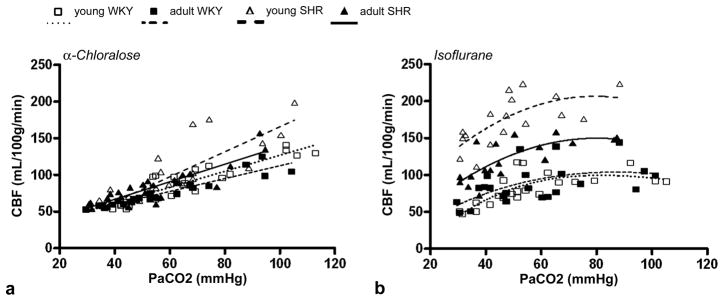
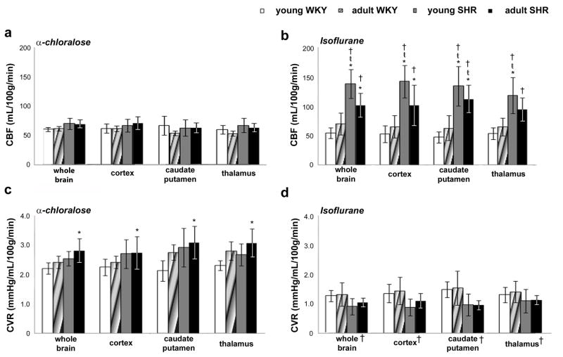
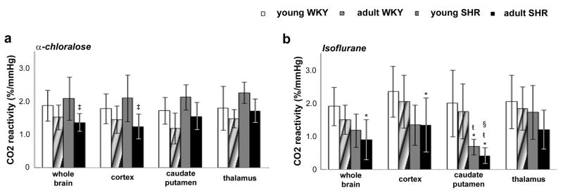
Hypertension afflicts 25% of the general population and over 50% of the elderly. In the present work, arterial spin labeling MRI was used to non-invasively quantify regional cerebral blood flow (CBF), cerebrovascular resistance and CO(2) reactivity in spontaneously hypertensive rats (SHR) and in normotensive Wistar Kyoto rats (WKY), at two different ages (3 months and 10 months) and under the effects of two anesthetics, α-chloralose and 2% isoflurane (1.5 MAC). Repeated CBF measurements were highly consistent, differing by less than 10% and 18% within and across animals, respectively. Under α-chloralose, whole brain CBF at normocapnia did not differ between groups (young WKY: 61 ± 3ml/100g/min; adult WKY: 62 ± 4ml/100g/min; young SHR: 70 ± 9ml/100g/min; adult SHR: 69 ± 8ml/100g/min), indicating normal cerebral autoregulation in SHR. At hypercapnia, CBF values increased significantly, and a linear relationship between CBF and PaCO(2) levels was observed. In contrast, 2% isoflurane impaired cerebral autoregulation. Whole brain CBF in SHR was significantly higher than in WKY rats at normocapnia (young SHR: 139 ± 25ml/100g/min; adult SHR: 104 ± 23ml/100g/min; young WKY: 55± 9ml/100g/min; adult WKY: 71 ± 19ml/100g/min). CBF values increased significantly with increasing CO(2); however, there was a clear saturation of CBF at PaCO(2) levels greater than 70mmHg in both young and adult rats, regardless of absolute CBF values, suggesting that isoflurane interferes with the vasodilatory mechanisms of CO(2). This behavior was observed for both cortical and subcortical structures. Under either anesthetic, CO(2) reactivity values in adult SHR were decreased, confirming that hypertension, when combined with age, increases cerebrovascular resistance and reduces cerebrovascular compliance.
Published by Elsevier Inc.
Figures

Similar articles
-
Regional cerebral blood flow and arterial blood volume and their reactivity to hypercapnia in hypertensive and normotensive rats.J Cereb Blood Flow Metab. 2014 Mar;34(3):408-14. doi: 10.1038/jcbfm.2013.197. Epub 2013 Nov 20. J Cereb Blood Flow Metab. 2014. PMID: 24252849 Free PMC article.
-
Intensity of halothane- and hypercapnia-induced cerebral hyperemia is strain-dependent in rats.Anesth Analg. 1996 Aug;83(2):359-65. doi: 10.1097/00000539-199608000-00027. Anesth Analg. 1996. PMID: 8694319
-
Cerebral angiography, blood flow and vascular reactivity in progressive hypertension.Neuroimage. 2015 May 1;111:329-37. doi: 10.1016/j.neuroimage.2015.02.053. Epub 2015 Feb 27. Neuroimage. 2015. PMID: 25731987 Free PMC article.
-
Impact of intravenous antihypertensive therapy on cerebral blood flow and neurocognition: a systematic review and meta-analysis.Br J Anaesth. 2025 Mar;134(3):713-726. doi: 10.1016/j.bja.2024.12.007. Epub 2025 Jan 20. Br J Anaesth. 2025. PMID: 39837698 Free PMC article.
-
Is the brain the essential in hypertension?Neuroimage. 2009 Sep;47(3):914-21. doi: 10.1016/j.neuroimage.2009.04.072. Epub 2009 May 4. Neuroimage. 2009. PMID: 19410005 Free PMC article. Review.
Cited by
-
Optimized analysis of blood flow and wall shear stress in the common carotid artery of rat model by phase-contrast MRI.Sci Rep. 2017 Jul 12;7(1):5253. doi: 10.1038/s41598-017-05606-4. Sci Rep. 2017. PMID: 28701695 Free PMC article.
-
A Review of Translational Magnetic Resonance Imaging in Human and Rodent Experimental Models of Small Vessel Disease.Transl Stroke Res. 2021 Feb;12(1):15-30. doi: 10.1007/s12975-020-00843-8. Epub 2020 Sep 16. Transl Stroke Res. 2021. PMID: 32936435 Free PMC article.
-
Comparing the effect of positioning on cerebral autoregulation during radical prostatectomy: a prospective observational study.J Clin Monit Comput. 2021 Aug;35(4):891-901. doi: 10.1007/s10877-020-00549-0. Epub 2020 Jun 20. J Clin Monit Comput. 2021. PMID: 32564173 Free PMC article.
-
The Effect of Chunghyul-Dan on Hyperventilation-Induced Carbon Dioxide Reactivity of the Middle Cerebral Artery in Normal Subjects: A Dose-Dependent Study.Evid Based Complement Alternat Med. 2017;2017:4567217. doi: 10.1155/2017/4567217. Epub 2017 Apr 20. Evid Based Complement Alternat Med. 2017. PMID: 28512500 Free PMC article.
-
Impaired CBF regulation and high CBF threshold contribute to the increased sensitivity of spontaneously hypertensive rats to cerebral ischemia.Neuroscience. 2014 Jun 6;269:223-31. doi: 10.1016/j.neuroscience.2014.03.031. Epub 2014 Mar 25. Neuroscience. 2014. PMID: 24680939 Free PMC article.
References
-
- Alson RL, Dusseau JW, Hutchins PM. Arteriolar and systemic autoregulatory responses during the development of hypertension in the spontaneously hypertensive rat. Proc Soc Exp Biol Med. 1985;180:62–71. - PubMed
-
- Alsop DC, Detre JA. Reduced transit-time sensitivity in noninvasive magnetic resonance imaging of human cerebral blood flow. J Cereb Blood Flow Metab. 1996;16:1236–1249. - PubMed
-
- Amenta F, Di Tullio MA, Tomassoni D. Arterial hypertension and brain damage--evidence from animal models (review) Clin Exp Hypertens. 2003;25:359–380. - PubMed
-
- Bakalova R, Matsuura T, Kanno I. Frequency dependence of local cerebral blood flow induced by somatosensory hind paw stimulation in rat under normo- and hypercapnia. Jpn J Physiol. 2001;51:201–208. - PubMed
-
- Barone FC, Price WJ, White RF, Willette RN, Feuerstein GZ. Genetic hypertension and increased susceptibility to cerebral ischemia. Neurosci Biobehav Rev. 1992;16:219–233. - PubMed
Publication types
MeSH terms
Substances
Grants and funding
LinkOut - more resources
Full Text Sources
Medical
