

Automatic segmentation of intracochlear anatomy in conventional CT
- PMID: 21708495
- PMCID: PMC3804019
- DOI: 10.1109/TBME.2011.2160262
Automatic segmentation of intracochlear anatomy in conventional CT
Abstract
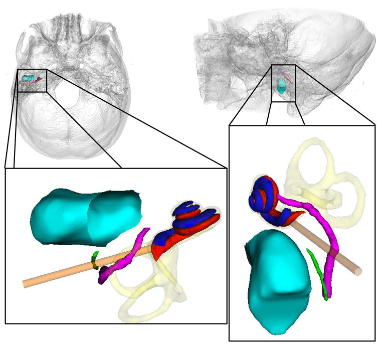
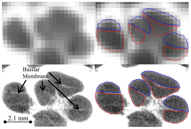
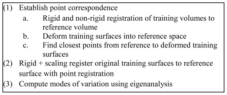
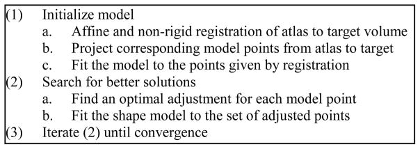
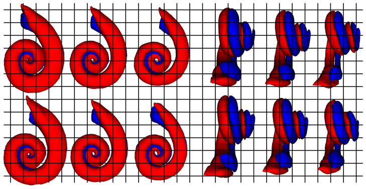
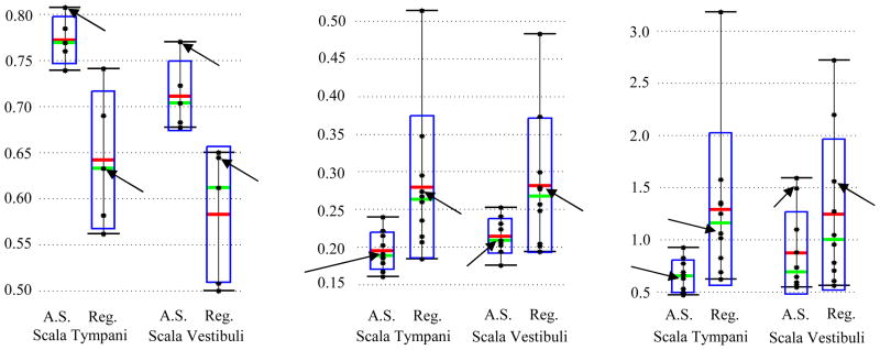
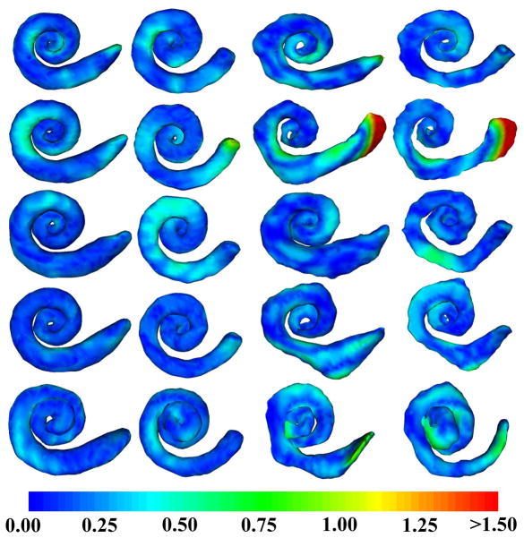
Cochlear implant surgery is a procedure performed to treat profound hearing loss. Clinical results suggest that implanting the electrode in the scala tympani, one of the two principal cavities inside the cochlea, may result in better hearing restoration. Segmentation of intracochlear cavities could thus aid the surgeon to choose the point of entry and angle of approach that maximize the likelihood of successful implant insertion, which may lead to more substantial hearing restoration. However, because the membrane that separates the intracochlear cavities is too thin to be seen in conventional in vivo imaging, traditional segmentation techniques are inadequate. In this paper, we circumvent this problem by creating an active shape model with micro CT (μCT) scans of the cochlea acquired ex vivo. We then use this model to segment conventional CT scans. The model is fitted to the partial information available in the conventional scans and used to estimate the position of structures not visible in these images. Quantitative evaluation of our method, made possible by the set of μCTs, results in Dice similarity coefficients averaging 0.75. Mean and maximum surface errors average 0.21 and 0.80 mm.
Figures

References
-
- Skinner MW, Holden TA, Whiting BR, et al. In vivo estimates of the position of advanced bionics electrode arrays in the human cochlea. Ann Otol Rhinol Laryngol Suppl. 2007 Apr;197:2–24. - PubMed
-
- Aschendorff A, Kromeier J, Klenzner T, Laszig R. Quality Control After Insertion of the Nucleus Contour and Contour Advance Electrode in Adults. Ear & Hearing. 2007 Apr;28:75S–79S. - PubMed
-
- Lehnhardt E, Laszig R. Specific surgical aspects of cochlear implant-soft surgery. Adv in coch Imp. 1994:228–9.
-
- James C, Albegger K, Battmer R, et al. Preservation of residual hearing with cochlear implantation: How and why. Acta Oto-Laryngologica. 2005;125(5):481–91. - PubMed
-
- Noble JH, Rutherford R, Labadie RF, Majdani O, Dawant BM. Modeling and segmentation of intra-cochlear anatomy in conventional CT. Proc of the SPIE conf on Med Imag. 2010;7623:762302.
Publication types
MeSH terms
Grants and funding
LinkOut - more resources
Full Text Sources
Other Literature Sources
Medical
