Review
. 2011 Sep 15;589(Pt 18):4413-21.
doi: 10.1113/jphysiol.2011.212712. Epub 2011 Jun 27.

Mitochondria: isolation, structure and function

Affiliations
Review

Mitochondria: isolation, structure and function

Martin Picard et al. J Physiol. .

Abstract

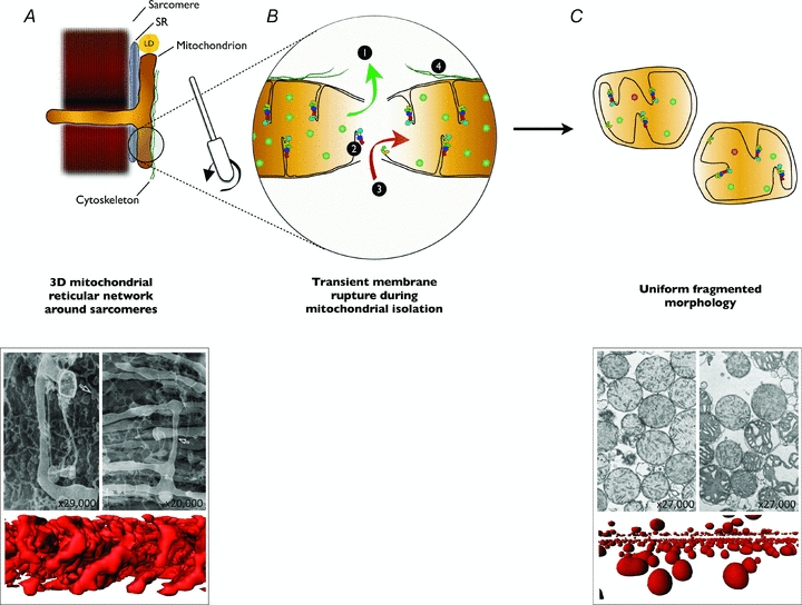
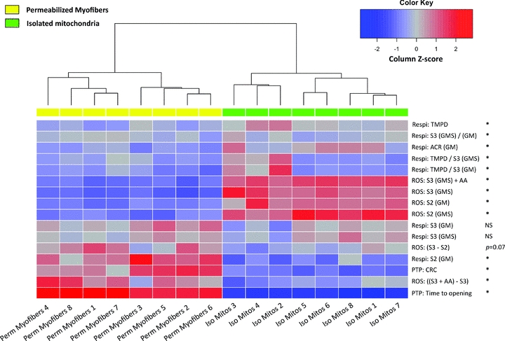
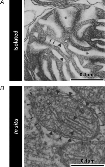
Mitochondria are complex organelles constantly undergoing processes of fusion and fission, processes that not only modulate their morphology, but also their function. Yet the assessment of mitochondrial function in skeletal muscle often involves mechanical isolation of the mitochondria, a process which disrupts their normally heterogeneous branching structure and yields relatively homogeneous spherical organelles. Alternatively, methods have been used where the sarcolemma is permeabilized and mitochondrial morphology is preserved, but both methods face the downside that they remove potential influences of the intracellular milieu on mitochondrial function. Importantly, recent evidence shows that the fragmented mitochondrial morphology resulting from routine mitochondrial isolation procedures used with skeletal muscle alters key indices of function in a manner qualitatively similar to mitochondria undergoing fission in vivo. Although these results warrant caution when interpreting data obtained with mitochondria isolated from skeletal muscle, they also suggest that isolated mitochondrial preparations might present a useful way of interrogating the stress resistance of mitochondria. More importantly, these new findings underscore the empirical value of studying mitochondrial function in minimally disruptive experimental preparations. In this review, we briefly discuss several considerations and hypotheses emerging from this work.


Figures

Figure 1
Figure 1. Mitochondrial isolation fragments mitochondria and alters mitochondrial function
A, in vivo and in situ, mitochondria exhibit a complex three-dimensional network arrangement around sarcomeres (Kirkwood et al. 1986; Ogata & Yamasaki, 1997). In addition, mitochondria maintain functional interactions with the sarcoplasmic reticulum (SR), lipid droplets (LD) (Goodman, 2008) and cytoskeleton (Saks et al. 2010). B, during mitochondrial isolation, muscle is homogenized using scissors and a Teflon pestle, which separates mitochondria and ruptures mitochondrial membranes. This transient rupture of mitochondrial membranes has four potential direct effects: allowing soluble mitochondrial molecules to escape the mitochondria, thereby diluting matrix components necessary for oxidative phosphorylation or reactive oxygen species detoxification (1); causing disruption of the inner mitochondrial membrane and the structural integrity of its residing supramolecular complexes (2); allowing soluble molecules from the homogenization medium to enter the mitochondria to alter aspects of mitochondrial function, or allowing the medium itself to enter the matrix causing swelling and dilution of matrix constituents (3); and disrupting functional interactions between mitochondria, SR, LDs and cytoskeleton (4). C, the end result of mitochondrial isolation is a relatively homogeneous population of spherical organelles with swollen morphology, disorganized cristae and diluted matrix content. Together, these effects result in altered functional characteristics of isolated mitochondria when compared to intact mitochondria from permeabilized myofibres. EM images reproduced from Ogata & Yamasaki (1997) and Hackenbrock (1968), with permission from Wiley and Rockefeller University Press, respectively. Three dimensional reconstructions are from MitoTracker-labelled confocal imaging of mitochondria in permeabilized myofibres or isolated mitochondria, as described in Picard et al. (2011).
Figure 2
Figure 2. Isolated mitochondria and permeabilized myofibres have different functional signatures
Heat map depicting results from an unsupervised hierarchical clustering analysis, whereby samples with the most similar functional signatures are grouped together. Functional signatures are defined here as the integration of functional measures of respiratory capacity (Respi) and H2O2 production (ROS) under various stimulatory conditions, as well as mitochondrial permeability transition pore (mPTP) responses to a Ca2+ challenge. The dendrogram above the heat map shows two primary clusters of similar compactness and exhibiting marked distinctiveness, corresponding to permeabilized myofibre and isolated mitochondrial preparations. The heat map analysis was performed with data obtained from mixed gastrocnemius muscles of Fisher 344/Brown Norway F1-hybrid rats (data reported in: Picard et al. 2011). To allow direct comparison of both preparations, all data are presented and normalized per international unit of citrate synthase activity, except for time to PTP opening expressed in seconds. The z-score for each value was computed from the mean for each measure. Measures which significantly differ (P < 0.05) between preparations are marked with an asterisk. Statistical analysis was performed using ‘R’ (R Foundation for Statistical Computing, version 2.12.1). List of abbreviations: TMPD, TMPD-driven complex IV respiration; S2, State 2 – Substrates-driven respiration; S3, State 3 – ADP-driven respiration with glutamate + malate (GM) or GM + succinate (GMS); ACR, acceptor control ratio; AA, antimycin A; CRC, calcium retention capacity; NS, not significant. Details of experimental procedures are reported in Picard et al. (2011).
Figure 3
Figure 3. Muscle mitochondrial ultrastructure is disrupted in isolated mitochondria (in vitro) compared to normal mitochondria in situ
Electron micrographs of mitochondria from skeletal muscle depicting the cristae morphology in isolated mitochondria (A) compared to mitochondria fixed in situ (B). Several differences are noted in isolated mitochondria, including: lower electron density of the matrix space, dysmorphic and irregular inner mitochondrial membrane cristae structure, absence of consistent contact sites near cristae junctions, and swelling of inter-membrane spaces. Images are from Schwerzmann et al. (1989) and are reproduced here with permission from the National Academy of Sciences of the USA.

References

    1. Adhihetty PJ, Ljubicic V, Menzies KJ, Hood DA. Differential susceptibility of subsarcolemmal and intermyofibrillar mitochondria to apoptotic stimuli. Am J Physiol Cell Physiol. 2005;289:C994–C1001. - PubMed
    1. Anderson EJ, Neufer PD. Type II skeletal myofibers possess unique properties that potentiate mitochondrial H2O2 generation. Am J Physiol Cell Physiol. 2006;290:C844–851. - PubMed
    1. Bakeeva LE, Chentsov Yu S, Skulachev VP. Mitochondrial framework (reticulum mitochondriale) in rat diaphragm muscle. Biochim Biophys Acta. 1978;501:349–369. - PubMed
    1. Benard G, Bellance N, James D, Parrone P, Fernandez H, Letellier T, Rossignol R. Mitochondrial bioenergetics and structural network organization. J Cell Sci. 2007;120:838–848. - PubMed
    1. Chance B, Williams GR. Respiratory enzymes in oxidative phosphorylation. VI. The effects of adenosine diphosphate on azide-treated mitochondria. J Biol Chem. 1956;221:477–489. - PubMed

MeSH terms

Substances

LinkOut - more resources