. 2011 Jul 4;208(7):1377-88.
doi: 10.1084/jem.20102065. Epub 2011 Jun 27.

B cell priming for extrafollicular antibody responses requires Bcl-6 expression by T cells

Affiliations

B cell priming for extrafollicular antibody responses requires Bcl-6 expression by T cells

Sau K Lee et al. J Exp Med. .

Abstract

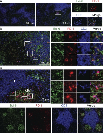
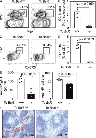
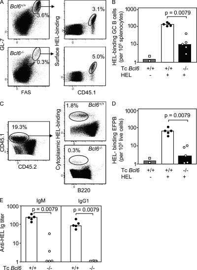
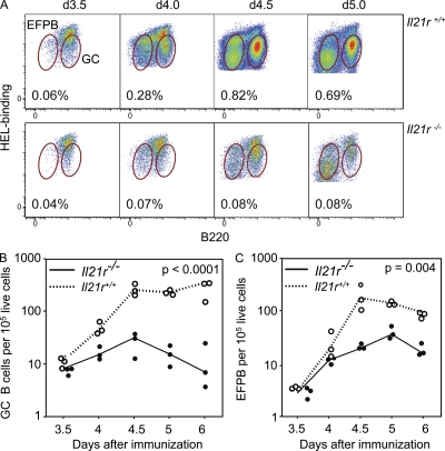
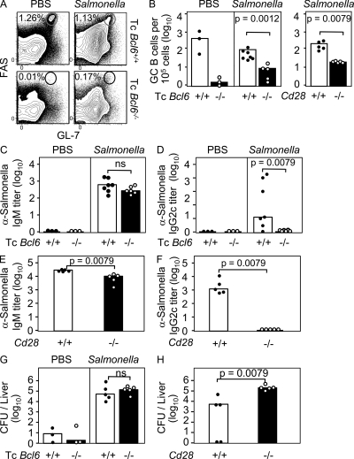
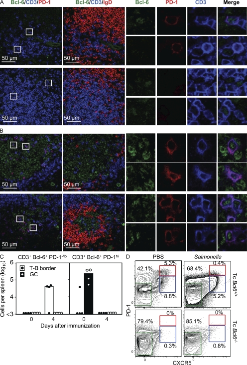
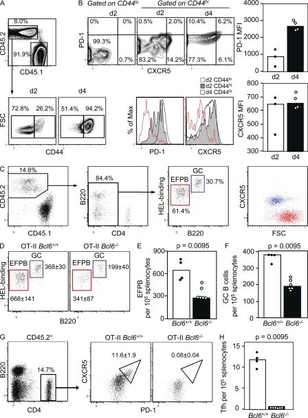
T follicular helper cells (Tfh cells) localize to follicles where they provide growth and selection signals to mutated germinal center (GC) B cells, thus promoting their differentiation into high affinity long-lived plasma cells and memory B cells. T-dependent B cell differentiation also occurs extrafollicularly, giving rise to unmutated plasma cells that are important for early protection against microbial infections. Bcl-6 expression in T cells has been shown to be essential for the formation of Tfh cells and GC B cells, but little is known about its requirement in physiological extrafollicular antibody responses. We use several mouse models in which extrafollicular plasma cells can be unequivocally distinguished from those of GC origin, combined with antigen-specific T and B cells, to show that the absence of T cell-expressed Bcl-6 significantly reduces T-dependent extrafollicular antibody responses. Bcl-6(+) T cells appear at the T-B border soon after T cell priming and before GC formation, and these cells express low amounts of PD-1. Their appearance precedes that of Bcl-6(+) PD-1(hi) T cells, which are found within the GC. IL-21 acts early to promote both follicular and extrafollicular antibody responses. In conclusion, Bcl-6(+) T cells are necessary at B cell priming to form extrafollicular antibody responses, and these pre-GC Tfh cells can be distinguished phenotypically from GC Tfh cells.


Figures

Figure 1.
Figure 1.
Bcl-6+ PD-1lo CD4+ cells are seen at the T–B border after SRBC immunization. (A–C) Immunofluorescence stains of spleen sections from C57BL/6 mice at 0 (A), 3 (B), and 7 d (C) after SRBC immunization. Boxed areas indicate the location of the zoomed-in images on the right, in the same order (from top to bottom). These data are representative of two independent experiments with four mice per experiment. F, follicle; T, T zone.
Figure 2.
Figure 2.
Reduced plasma cell responses to NP-KLH in the absence of T cell–expressed Bcl-6. (A–D) Representative flow cytometric contour plots (A and C) and quantification (B and D) of GC B cells identified as B220+ PNAhi splenocytes (A and B) and Tfh cells identified as CXCR5hi PD-1hi (gated on CD4+ cells; C and D) from 80% Bcl6−/− CD45.2:20% Cd28−/− CD45.1 (i.e., effector T cells lack Bcl-6; Tc Bcl6−/−) or control 80% Bcl6+/+ CD45.2:20% Cd28−/− CD45.1 (Tc Bcl6+/+) fetal liver chimeric mice immunized 7 d earlier with NP-KLH. (E) NP-specific IgG1 and NP-specific IgM antibodies in the same chimeric mice. (F) Photomicrographs of spleen sections stained with B220 (follicles; blue) and IgG1 (plasma cells; brown). These data are representative of two independent experiments with five mice per group in each cohort.
Figure 3.
Figure 3.
Development of HEL-specific EFPBs cells is greatly impaired in the absence of T cell–expressed Bcl-6. (A–D) Representative flow cytometric contour plots (A and C) and quantification (B and D) of SWHEL GC B cells identified as B220+ GL-7+ Fas+ HEL-binding CD45.1 cells (A and B) and EFPBs identified as CD45.1+ B220lo intracellular HEL-binding cells (C and D) in CD45.2 chimeric mice sufficient (Tc Bcl6+/+) or deficient (Tc Bcl6−/−) in Bcl-6–expressing T cells 4.5 d after adoptive transfer of SWHEL B cells and HEL-SRBC immunization. Control (HEL) mice received SWHEL B cells and were immunized with SRBCs that had not been conjugated to HEL. (E) HEL-specific antibody titers in the same HEL-SRBC–immunized chimeric mice. These data are representative of two independent experiments with five mice per group immunized with HEL-SRBC and two mice per group in the HEL-only control group for each independent experiment.
Figure 4.
Figure 4.
IL-21 influences GC and EFPB responses from the early stages of B cell activation. (A) Representative flow cytometric dot plots showing HEL binding versus B220 staining of Il21r+/+ or Il21r−/− CD45.2 SWHEL B cells at the indicated days after transfer into congenic (CD45.1) C57BL/6 recipients immunized with HEL2x-SRBC at the time of transfer. The gates in each plot are drawn around GC B cells (B220hi; right) and EFPBs (B220lo; left). Numbers in plots indicate the percentage of HEL-binding CD45.2 SWHEL cells out of total splenocytes. (B and C) Number of SWHEL GC B cells (B) and EFPBs (C) in the same mice using the gates shown in A. These data are representative of two independent experiments with three mice per group per time point. The p-values were calculated using two-way analysis of variance that interrogates the variance over the entire time course between the two groups of mice receiving either Il21r+/+ or Il21r−/− SWHEL B cells.
Figure 5.
Figure 5.
IgG2c extrafollicular antibody responses to S. enterica are impaired in the absence of T cell–expressed Bcl-6. (A) Representative flow cytometric plots showing Fas versus GL-7 stains from spleens of 80% Bcl6−/− CD45.2:20% Cd28−/− CD45.1 (Tc Bcl6−/−) chimeras or control 80% Bcl6+/+ CD45.2:20% Cd28−/− CD45.1 (Tc Bcl6+/+) fetal liver chimeric mice injected 7 d previously with S. enterica; oval gates identify GC B cells. (B) Bar graphs show the number of splenic GC B cells in the same mice (left) and a group of Cd28−/− and control Cd28+/+ mice infected with S. enterica at the same time (right). (C–F) S. enterica–specific IgM (C and E) and IgG2c (D and F) antibody titers 7 d after S. enterica infection in the same fetal liver chimeric mice as in A (C and D) or Cd28−/− and Cd28+/+ mice (E and F). (G and H) S. enterica CFUs in livers 12 d after infection of the chimeras (G) and nonchimeric Cd28−/− and Cd28+/+ mice (H). These data are representative of three independent experiments with three mice per PBS-only control groups and five to seven mice per S. enterica–infected groups.
Figure 6.
Figure 6.
Bcl-6+ T cells induced by S. enterica infection are PD-1lo and locate to the T–B border. (A and B) Immunofluorescence stains of spleen sections from C57BL/6 immunized 4 (A) or 35 d (B) previously with S. enterica. Boxed areas indicate the location of the zoomed-in images on the right, in the same order (from top to bottom). (C) Quantification of CD3+ Bcl-6+ PD-1lo (left) and CD3+ Bcl-6+ PD-1hi (right) cells in the T–B border and GCs at the indicated times after immunizations. (D) Representative flow cytometric plots showing PD-1 versus CXCR5 expression on effector/memory CD4+ CD44hi cells from 80% Bcl6−/− CD45.2:20% Cd28−/− CD45.1 (Tc Bcl6−/−) chimeras or control 80% Bcl6+/+ CD45.2:20% Cd28−/− CD45.1 (Tc Bcl6+/+) fetal liver chimeric mice injected 7 d previously with S. enterica (green gate, non-Tfh effectors; blue gate, PD-1lo Tfh cells; and red gate, PD-1hi Tfh cells). These data are representative of two independent experiments with four mice per group in each cohort.
Figure 7.
Figure 7.
Development of HEL-specific GC and EFPBs is impaired in the absence of OVA-specific T cells expressing Bcl-6. Naive OT-II Bcl6+/+ or OT-II Bcl6−/− with or without SWHEL B cells were transferred i.v. 4 h apart into Cd28−/− CD45.1 recipient mice, which were then immunized with HEL-OVA and/or OVA in alum i.p. (A and B) CXCR5 versus PD-1 phenotype of OT-II Bcl6+/+ cells on day 2 or 4 after transfer into mice immunized with OVA in alum. Gating strategy to identify naive versus activated donor OT-II cells on day 2 or 4 after transfer (A) and representative flow cytometric plots, histograms, and bar graphs showing PD-1/ CXCR5 expression on naive (CD44lo) and activated (CD44hi) OT-II cells on either day 2 or 4 after adoptive cell transfer and immunization, analyzed on the same day (B). (C) Representative flow cytometric plots showing the gating strategy to identify donor-origin SWHEL GC B cells (CD45.2 B220hi HEL-bindinghi CXCR5hi small forward scatter [FSC]) and EFPBs (CD45.2 B220lo HEL-bindinglo CXCR5lo large forward scatter) in the spleens of Cd28−/− CD45.1 recipient mice 5.5 d after adoptive transfer. (D–F) Flow cytometric profiles (D) and quantification of SWHEL EFPBs (E) and GC B cells (F) in the same recipient mice 5.5 d after immunization with HEL-OVA. (G and H) Representative flow cytometric plot showing gating strategy (G) and quantification (H) of donor-origin Tfh cells identified as CXCR5hi PD-1hi CD45.2 (gated on CD4+ cells) after transfer of OT-II Bcl6+/+ or OT-II Bcl6−/− T cells. These data are representative of two independent experiments with three to six mice per group.

Similar articles

Cited by

References

    1. Cannons J.L., Qi H., Lu K.T., Dutta M., Gomez-Rodriguez J., Cheng J., Wakeland E.K., Germain R.N., Schwartzberg P.L. 2010. Optimal germinal center responses require a multistage T cell:B cell adhesion process involving integrins, SLAM-associated protein, and CD84. Immunity. 32:253–265 10.1016/j.immuni.2010.01.010 - DOI - PMC - PubMed
    1. Chan T.D., Gatto D., Wood K., Camidge T., Basten A., Brink R. 2009. Antigen affinity controls rapid T-dependent antibody production by driving the expansion rather than the differentiation or extrafollicular migration of early plasmablasts. J. Immunol. 183:3139–3149 10.4049/jimmunol.0901690 - DOI - PubMed
    1. Cunningham A.F., Khan M., Ball J., Toellner K.M., Serre K., Mohr E., MacLennan I.C. 2004. Responses to the soluble flagellar protein FliC are Th2, while those to FliC on Salmonella are Th1. Eur. J. Immunol. 34:2986–2995 10.1002/eji.200425403 - DOI - PubMed
    1. Cunningham A.F., Gaspal F., Serre K., Mohr E., Henderson I.R., Scott-Tucker A., Kenny S.M., Khan M., Toellner K.M., Lane P.J., MacLennan I.C. 2007. Salmonella induces a switched antibody response without germinal centers that impedes the extracellular spread of infection. J. Immunol. 178:6200–6207 - PubMed
    1. Cyster J.G., Ansel K.M., Reif K., Ekland E.H., Hyman P.L., Tang H.L., Luther S.A., Ngo V.N. 2000. Follicular stromal cells and lymphocyte homing to follicles. Immunol. Rev. 176:181–193 10.1034/j.1600-065X.2000.00618.x - DOI - PubMed

Publication types

MeSH terms