. 2011 Jul 4;208(7):1459-71.
doi: 10.1084/jem.20102510. Epub 2011 Jun 27.

Severe lung fibrosis requires an invasive fibroblast phenotype regulated by hyaluronan and CD44

Affiliations

Severe lung fibrosis requires an invasive fibroblast phenotype regulated by hyaluronan and CD44

Yuejuan Li et al. J Exp Med. .

Abstract

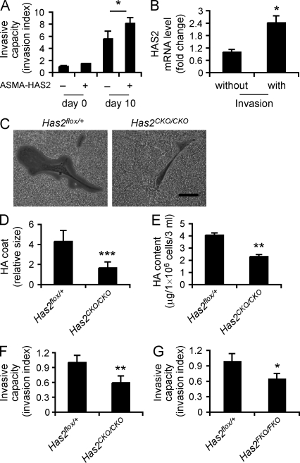
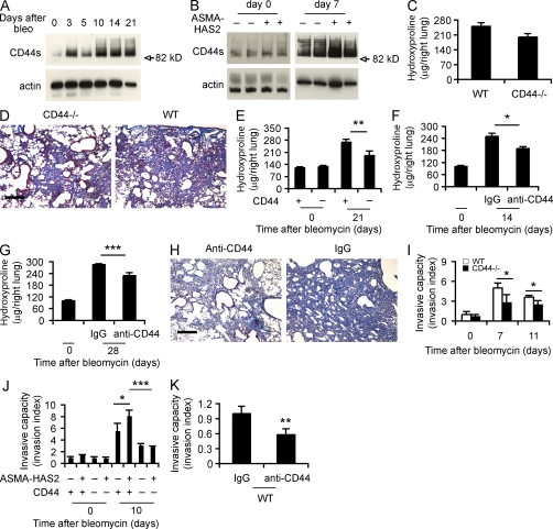
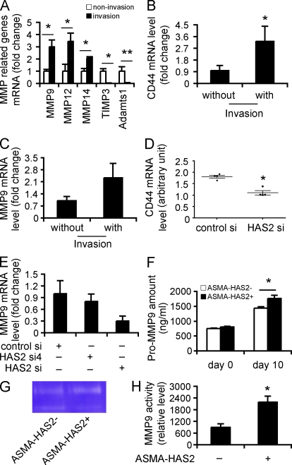
Tissue fibrosis is a major cause of morbidity, and idiopathic pulmonary fibrosis (IPF) is a terminal illness characterized by unremitting matrix deposition in the lung. The mechanisms that control progressive fibrosis are unknown. Myofibroblasts accumulate at sites of tissue remodeling and produce extracellular matrix components such as collagen and hyaluronan (HA) that ultimately compromise organ function. We found that targeted overexpression of HAS2 (HA synthase 2) by myofibroblasts produced an aggressive phenotype leading to severe lung fibrosis and death after bleomycin-induced injury. Fibroblasts isolated from transgenic mice overexpressing HAS2 showed a greater capacity to invade matrix. Conditional deletion of HAS2 in mesenchymal cells abrogated the invasive fibroblast phenotype, impeded myofibroblast accumulation, and inhibited the development of lung fibrosis. Both the invasive phenotype and the progressive fibrosis were inhibited in the absence of CD44. Treatment with a blocking antibody to CD44 reduced lung fibrosis in mice in vivo. Finally, fibroblasts isolated from patients with IPF exhibited an invasive phenotype that was also dependent on HAS2 and CD44. Understanding the mechanisms leading to an invasive fibroblast phenotype could lead to novel approaches to the treatment of disorders characterized by severe tissue fibrosis.


Figures

Figure 1.
Figure 1.
ASMA-HAS2 transgenic mice accumulate HA and show increased mortality after bleomycin challenge. (A) Distribution of HA in lungs of ASMA-HAS2+ and littermate control mice (ASMA-HAS2) was determined by immunohistochemical staining of HA with biotin-HABP. Representative sections from five bleomycin-treated samples and three controls are shown. The specificity of the staining was determined by preincubating tissue samples with 10 U/ml streptomyces hyaluronidase for 2 h at room temperature (not depicted). Bar, 50 µm. (B and C) HA concentration in lung tissue (B) and BALF (C) from ASMA-HAS2+ mice and control mice (ASMA-HAS2) at different times after bleomycin treatment (n = 5–7; *, P < 0.05; **, P < 0.01). (D and E) Murine HAS2 (mHAS2; D and E) and human HAS2 (hHAS2; E) mRNA expression levels in ASMA-HAS2+ mice and controls (ASMA-HAS2) at various time points after bleomycin treatment were measured using real-time PCR (n = 3–5; **, P < 0.01; ***, P < 0.001). (B–E) Error bars indicate mean ± SEM. (F) ASMA-HAS2+ and control mouse lung injury was induced after intratracheal inoculation with the indicated dose of bleomycin. Percentages of surviving mice were plotted over a 21-d period (for 1.75 U/kg: n = 8 per group, P = 0.14; for 2.5 U/kg: n = 16 per group, P = 0.05; for 5.0 U/kg: n = 9 per group, P < 0.05; red line, ASMA-HAS2; blue line, ASMA-HAS2+). (A–F) Experiments were performed three times.
Figure 2.
Figure 2.
ASMA-HAS2 transgenic mice exhibit increased collagen content in lungs after bleomycin treatment. (A) Lung sections of ASMA-HAS2+ and transgene negative controls 0, 14, and 28 d after bleomycin instillation were stained using Masson’s trichrome method. Representative images of the staining are shown (n = 6–7). (B) Lung tissues from ASMA-HAS2+ and controls on days 0, 14, 21, and 28 after bleomycin treatment were collected and assayed for collagen content using the hydroxyproline method (n = 6–7 per group; *, P < 0.05; ****, P < 0.0001). The experiments were performed three times. Error bars indicate mean ± SEM. (C and D) Immunohistochemical (C) and immunofluorescent analysis (D) of ASMA and HA in lung sections of ASMA-HAS2+ and control mice 14 d after bleomycin treatment. Representative images of the staining are shown (n = 6–7). (C) DAB staining is shown. (D) Immunofluorescence staining is shown. (E) Representative images of IPF patients’ and control lung tissues showing ASMA staining and similar fibrotic changes to bleomycin-induced lung fibrosis. Bars: (A and E) 200 µm; (C and D) 50 µm.
Figure 3.
Figure 3.
Targeted deletion of HAS2 in mesenchymal cells inhibits lung fibrosis and myofibroblast accumulation. (A) HA content in lung tissue from Has2FKO/FKO and Has2flox/+ mice on day 14 after bleomycin treatment (n = 3–8 per group; *, P < 0.05). (B) Lung tissues from Has2FKO/FKO and control Has2flox/+ mice on days 0, 14, and 21 after bleomycin treatment were collected and assayed for collagen content using the hydroxyproline method (n = 4–11 per group; *, P < 0.05). (A and B) Error bars indicate mean ± SEM. (C) Lung sections of Has2FKO/FKO and Has2flox/+ mice on days 0 and 14 after bleomycin instillation were stained using Masson’s trichrome method and counterstained with hematoxylin (n = 8 in each group). (D) Double staining of HA and ASMA in bleomycin-treated Has2FKO/FKO and control Has2flox/+ mouse lung sections on days 0 and 14 after bleomycin instillation. The experiments were performed three times. Bars, 200 µm.
Figure 4.
Figure 4.
Fibroblast invasive capacity is dependent on HAS2. (A) The spontaneous Matrigel-invading capacity of fibroblasts from bleomycin-treated (10 d) and saline-treated ASMA-HAS2+ and littermate control mice lungs was determined. Data are shown as the index of invasion value of the fibroblasts with or without bleomycin treatment over littermate control fibroblasts without bleomycin challenge (n = 4 per group; *, P < 0.05). (B) mRNA relative levels of HAS2 in invasive and noninvasive fibroblasts isolated from bleomycin-treated (11 d) WT mouse lungs were determined using real-time PCR (n = 5; *, P < 0.05). (C) Phase-contrast photomicrographs of the pericellular matrices (HA coat) in Has2CKO/CKO fibroblasts compared with those in Has2flox/+ fibroblasts. Bar, 50 µm. (A–C) Experiments were performed three times. (D) Relative thickness of HA coat was calculated in 20 randomly selected cells using ImageJ (n = 10; ***, P < 0.001). Data represent one of two independent experiments. (E) HA content in cultured media of Has2flox/+ and Has2CKO/CKO fibroblasts was measured using the HA-ELISA assay (n = 3; **, P < 0.01). The experiments were performed three times. (F) Comparison of the invasive capacity between Has2flox/+ and Has2CKO/CKO fibroblasts. Data are shown as the invasion index of Has2CKO/CKO fibroblasts over Has2flox/+ fibroblasts. They are representative of three independent experiments (n = 3; **, P < 0.01). (G) Comparison of the invasive capacity between fibroblasts from bleomycin-treated Has2flox/+ and Has2FKO/FKO mice. Data are shown as the invasion index of Has2FKO/FKO fibroblasts over Has2flox/+ fibroblasts (n = 3; *, P < 0.05). The experiments were performed three times. (A, B, and D–G) Error bars indicate mean ± SEM.
Figure 5.
Figure 5.
CD44 regulates lung fibrosis and fibroblast invasive capacity. (A) Western blot analysis of CD44 expression using KM114 anti-CD44 antibodies in WT lung tissues at the indicated times after bleomycin treatment. Samples loaded at each time point were the mixture of equal amounts of three samples collected per time point. β-Actin was used as a loading control. CD44 standard form (82.0 kD) is indicated. (B) Immunoblot of CD44 in ASMA-HAS2+ (+) and control (−) mouse lung tissues on days 0 and 7 after bleomycin treatment. (C) Lung tissues from CD44-null and WT mice on day 21 after bleomycin treatment were collected and assayed for collagen content using the hydroxyproline method (n = 14–17 per group). (A–C) The experiments were performed three times. (D) Lung sections of WT and CD44-null mice on day 21 after bleomycin instillation were stained using Masson’s trichrome method. Representative images of the staining are shown (n = 5–6). The experiment was repeated twice. (E) Hydroxyproline content on days 0 and 21 after bleomycin treatment was analyzed in ASMA-HAS2+/CD44+/+ and ASMA-HAS2+/CD44−/− mice (n = 7–8 per group; **, P < 0.01). (F) Neutralizing anti-CD44 antibodies were instilled i.p. 12 h before and 5 d after bleomycin treatment in ASMA-HAS2+ mice. Lungs were analyzed for hydroxyproline content on day 14 after bleomycin instillation (n = 5–8 per group; *, P < 0.05). (G) Anti-CD44 neutralizing antibodies were instilled i.p. on days 7, 14, and 21 after bleomycin treatment, and lungs were analyzed for hydroxyproline content at day 28 (n = 6–9 per group; ***, P < 0.001). (E–G) The experiments were performed three times. (H) Lung sections of the mice described in G were stained using Masson’s trichrome method. Representative images of the staining are shown. (I) The spontaneous Matrigel-invading capacity of fibroblasts from bleomycin-treated (7 and 11 d) and saline-treated WT C57BL/6J and CD44-null mouse lungs was determined. Data are shown as the index of invasion value of the fibroblasts with or without bleomycin treatment over WT fibroblasts without bleomycin challenge (n = 4 per group; *, P < 0.05). (J) Invasive capacity of mesenchymal cells from ASMA-HAS2, ASMA-HAS2+, ASMA-HAS2/CD44−/−, and ASMA-HAS2+/CD44−/− mouse lungs with or without bleomycin challenge was compared. Data are shown as the index of invasion value of the fibroblasts with or without bleomycin treatment over ASMA-HAS2 fibroblasts without bleomycin challenge (n = 4 per group; *, P < 0.05; ***, P < 0.001). (K) Invasion of bleomycin-treated WT mouse lung fibroblasts with (anti-CD44) or without (IgG) neutralizing CD44 antibody incubation (n = 4 per group; **, P < 0.01). (I–K) The experiments were repeated three times. (C, E–G, and I–K) Error bars indicate mean ± SEM. Bars, 200 µm.
Figure 6.
Figure 6.
HAS2 and CD44 are required for human lung fibroblast invasion. (A) Invasive capacity of human fibroblasts from normal subjects (NHF; n = 5) and IPF patients (n = 9). Results are for five separate experiments and are expressed as the invasion index of the IPF fibroblasts over the normal fibroblasts (*, P < 0.05). (B) Representative images of invasive IPF fibroblasts and normal fibroblasts. (C) Relative HAS2 mRNA levels of invasive and noninvasive IPF fibroblasts were determined using real-time PCR (n = 7; *, P < 0.05). The experiments were repeated two times. (D) 48 h after transfection with HAS2 siRNA (HAS2 si) and control siRNA (control si), photomicrographs demonstrating the effects of HAS2 si on fibroblast cellular surface HA, photomicrographs demonstrating the effects of HAS2 si on HA coat formation, and images of invasive HAS2 siRNA– and control siRNA–transfected fibroblasts are shown. (E) 48 h after HAS2 and control siRNA transfection, equal numbers of fibroblasts from normal donors (n = 2) and IPF patients (n = 3) were loaded into invasion chambers and incubated for another 24 h. Invasive cells were counted. Data are shown as the invasion index of HAS2 siRNA–transfected normal, IPF fibroblasts and control siRNA–transfected IPF fibroblasts over control siRNA–transfected normal fibroblasts (**, P < 0.01). (F) After 20 min of incubation with anti-CD44 neutralizing or isotype-matched control IgG antibody, fibroblasts from normal donors (n = 3) and IPF patients (n = 6) were subjected to the invasion assay. Data are depicted as the invasion index (**, P < 0.01). (D–F) The experiments were repeated three times. (A, C, E, and F) Error bars indicate mean ± SEM. Bars: (B and D [top and bottom]) 200 µm; (D, middle) 100 µm.
Figure 7.
Figure 7.
HAS2 promotes fibroblast invasion by regulating CD44 and MMP expression and function. (A and B) RNA was extracted from invasive ASMA-HAS2+ fibroblasts, and 84 genes were analyzed by using a specialized qRT-PCR array for extracellular matrix synthesizing and degrading enzymes. RNAs from the fibroblasts that penetrated the filter in the absence of Matrigel were used as control. Representative genes up- or down-regulated in invasive ASMA-HAS2+ fibroblasts are shown (n = 5; *, P < 0.05; **, P < 0.01). (B) CD44 mRNA expression in invasive fibroblasts versus noninvasive fibroblasts from bleomycin-treated ASMA-HAS2+ lungs (n = 5; *, P = 0.05). (C) MMP9 mRNA expression in invasive IPF fibroblasts was compared with IPF fibroblasts that penetrated the filters in the absence of Matrigel by using real-time PCR (n = 5). The experiments were performed twice. (D) CD44 mRNA expression in HAS2 siRNA (HAS2 si)–transfected fibroblasts and control siRNA (control si) transfectants was determined using a microarray assay. The horizontal bars indicate the median expression values (n = 4; *, P < 0.05). (E) 48 h after HAS2 siRNA and control siRNA transfection, fibroblasts were cultured on Matrigel for an additional 6 h. mRNA was then extracted, and MMP9 mRNA expression was measured using real-time PCR. Data shown represent one of two separate experiments. (F) Fibroblasts from ASMA-HAS2 and ASMA-HAS2+ mice were cultured on Matrigel for 96 h, and pro-MMP9 protein in the media was measured using a pro-MMP9 ELISA kit (n = 3–7 per group; *, P < 0.05). The experiments were performed three times. (G) The media described in F was concentrated 10× using Microsep centrifugal devices. Equal amounts of protein were subjected to gelatin zymography for MMP9 activity. A representative image is shown. The experiments were repeated two times. (H) Quantification analysis of gelatin zymography for MMP9 activity results by using ImageJ software (n = 4–5 per group; *, P < 0.05). (A–F and H) Error bars indicate mean ± SEM.

References

    1. Acharya P.S., Majumdar S., Jacob M., Hayden J., Mrass P., Weninger W., Assoian R.K., Puré E. 2008. Fibroblast migration is mediated by CD44-dependent TGF beta activation. J. Cell Sci. 121:1393–1402 10.1242/jcs.021683 - DOI - PubMed
    1. Adamson I.Y., Bowden D.H. 1974. The pathogenesis of bleomycin-induced pulmonary fibrosis in mice. Am. J. Pathol. 77:185–197 - PMC - PubMed
    1. Ando H., Kubin T., Schaper W., Schaper J. 1999. Cardiac microvascular endothelial cells express alpha-smooth muscle actin and show low NOS III activity. Am. J. Physiol. 276:H1755–H1768 - PubMed
    1. Arch R., Wirth K., Hofmann M., Ponta H., Matzku S., Herrlich P., Zöller M. 1992. Participation in normal immune responses of a metastasis-inducing splice variant of CD44. Science. 257:682–685 10.1126/science.1496383 - DOI - PubMed
    1. ATS/ERS 2000. American Thoracic Society. Idiopathic pulmonary fibrosis: diagnosis and treatment. International consensus statement. American Thoracic Society (ATS), and the European Respiratory Society (ERS). Am. J. Respir. Crit. Care Med. 161:646–664 - PubMed

Publication types

MeSH terms