. 2011 Aug 12;286(32):28608-18.
doi: 10.1074/jbc.M111.225839. Epub 2011 Jun 27.

Vesicle-associated membrane protein-2 (VAMP2) mediates cAMP-stimulated renin release in mouse juxtaglomerular cells

Affiliations

Vesicle-associated membrane protein-2 (VAMP2) mediates cAMP-stimulated renin release in mouse juxtaglomerular cells

Mariela Mendez et al. J Biol Chem. .

Abstract

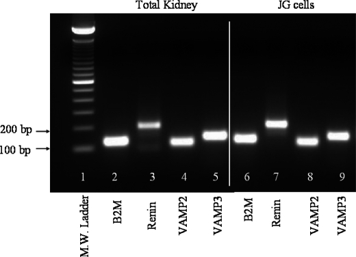
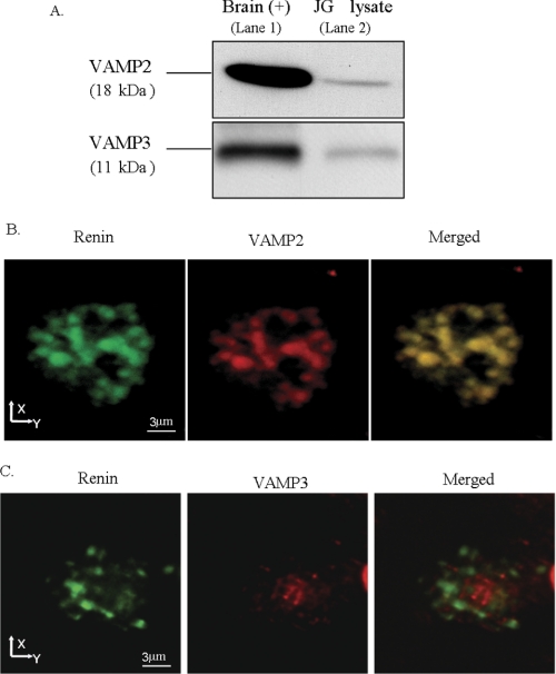
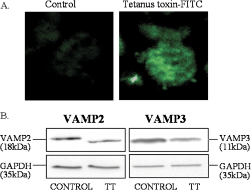
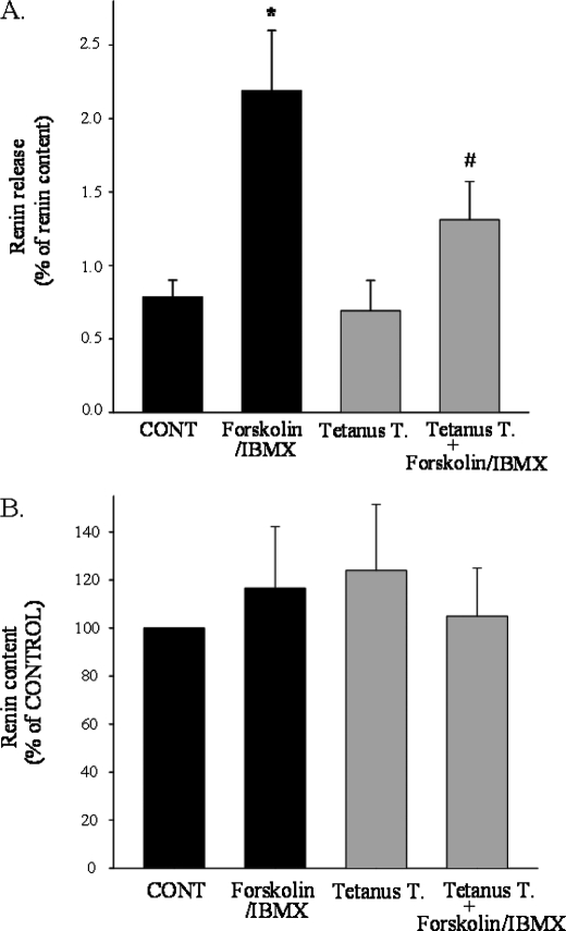
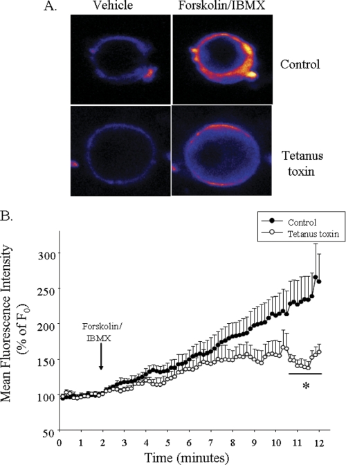
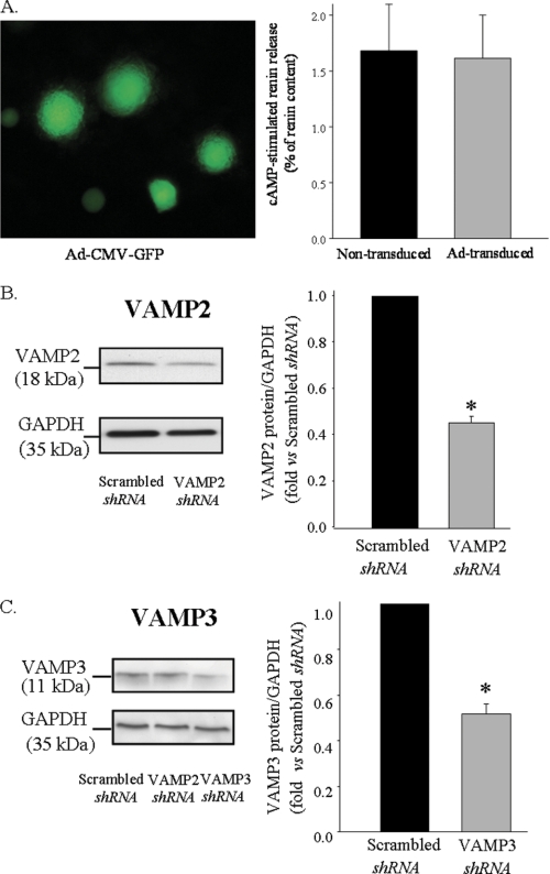
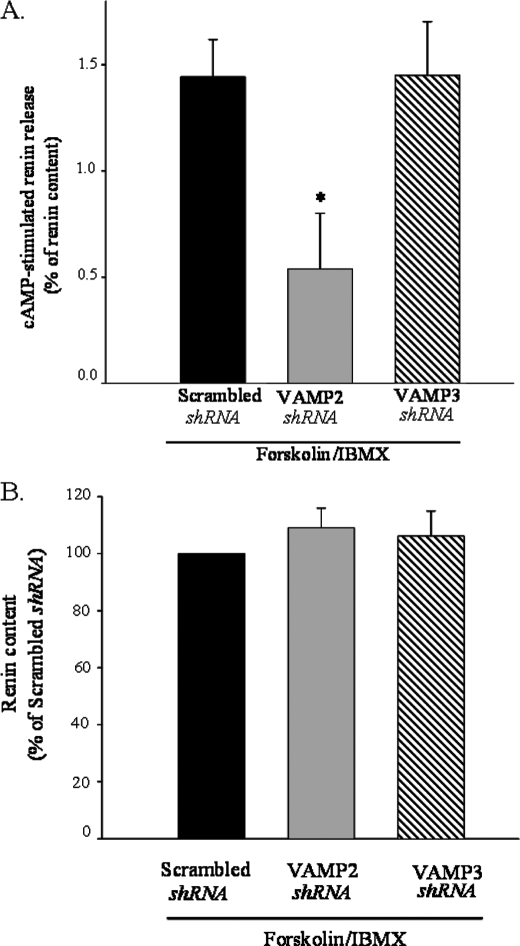
Renin is essential for blood pressure control. Renin is stored in granules in juxtaglomerular (JG) cells, located in the pole of the renal afferent arterioles. The second messenger cAMP stimulates renin release. However, it is unclear whether fusion and exocytosis of renin-containing granules is involved. In addition, the role of the fusion proteins, SNAREs (soluble N-ethylmaleimide-sensitive factor attachment proteins), in renin release from JG cells has not been studied. The vesicle SNARE proteins VAMP2 (vesicle associated membrane protein 2) and VAMP3 mediate cAMP-stimulated exocytosis in other endocrine cells. Thus, we hypothesized that VAMP2 and/or -3 mediate cAMP-stimulated renin release from JG cells. By fluorescence-activated cell sorting, we isolated JG cells expressing green fluorescent protein and compared the relative abundance of VAMP2/3 in JG cells versus total mouse kidney mRNA by quantitative PCR. We found that VAMP2 and VAMP3 mRNA are expressed and enriched in JG cells. Confocal imaging of primary cultures of JG cells showed that VAMP2 (but not VAMP3) co-localized with renin-containing granules. Cleavage of VAMP2 and VAMP3 with tetanus toxin blocked cAMP-stimulated renin release from JG cells by ~50% and impaired cAMP-stimulated exocytosis by ~50%, as monitored with FM1-43. Then we specifically knocked down VAMP2 or VAMP3 by adenoviral-mediated delivery of short hairpin silencing RNA. We found that silencing VAMP2 blocked cAMP-induced renin release by ~50%. In contrast, silencing VAMP3 had no effect on basal or cAMP-stimulated renin release. We conclude that VAMP2 and VAMP3 are expressed in JG cells, but only VAMP2 is targeted to renin-containing granules and mediates the stimulatory effect of cAMP on renin exocytosis.


Figures

FIGURE 1.
FIGURE 1.
Representative image of quantitative PCR products for VAMP2 and VAMP3. PCR products were resolved in an agarose gel stained with ethidium bromide (n = 3). Lane 1 is molecular weight ladder. Lanes 2–5 are PCR products from total kidney homogenate illustrating B2M, renin, VAMP2, and VAMP3, respectively. Lanes 6–9 correspond to PCR products from freshly isolated JG cells B2M, renin, VAMP2, and VAMP3, respectively. Some lanes not relevant to the current study have been intentionally cut out of the picture.
FIGURE 2.
FIGURE 2.
Expression and subcellular localization of VAMP2 and VAMP3 in JG cells. A, a representative Western blot shows expression of VAMP2 (18 kDa) and VAMP3 (11 kDa) in a JG cell lysate. The top panel is VAMP2, and the bottom panel is VAMP3. Lane 1 is brain homogenate (2.5 μg) used as a positive control, and lane 2 is JG cell lysate (7.5 μg) (n = 4). B, immunofluorescence and confocal microscopy of VAMP2 and renin on a single mouse JG cell are shown. The left panel shows a representative image from five preparations of a single JG cell labeled with an antibody against renin (green); 32 confocal slices (z-step 0.3 μm) were stacked into one image projection. Large renin-containing granules that range in size from 0.8 to 1.5 μm can be observed. The middle panel shows VAMP2 labeling (red) in the same cell. The right panel shows a merged image illustrating co-localization of renin with VAMP2 as illustrated by a yellow-orange color (n = 5 different preparations). Bar, 3 μm. C, immunofluorescence and confocal microscopy of VAMP3 and renin on a single mouse JG cell are shown. The left panel shows an individual JG cell immunolabeled with renin (green); the middle panel (red) is the same JG cell labeled with VAMP3 antibody; the right panel is the merged image. No co-localization was observed between small VAMP3-labeled vesicles and renin granules.
FIGURE 3.
FIGURE 3.
Internalization and cleavage of VAMP2 and VAMP3 by tetanus toxin. A, tetanus toxin is efficiently internalized in intact JG cells. JG cells were incubated for 19 h with either vehicle (Control; left panel) or FITC-labeled receptor binding domain of tetanus toxin (C-fragment; List Biological Laboratories) (right panel). After fixation and mounting, incorporated fluorescence was analyzed by confocal fluorescence imaging. B, representative Western blots show efficient cleavage of VAMP2 and VAMP3 by tetanus toxin. The left panel is VAMP2, and the right panel is VAMP3 protein expression. JG cells were incubated with either vehicle (left lane in both panels) or tetanus toxin (right lane in both panels). After treatment, JG cells were lysed, and SDS-PAGE was resolved in 12% polyacrylamide gel and transferred to PVDF membranes, which were subsequently blotted with VAMP2 or VAMP3 antibodies. Note that a decrease in VAMP2 and VAMP3 band intensity reflects efficient cleavage by tetanus toxin. Membranes were re-blotted with an antibody against GAPDH as an internal loading control (Mr ∼ 35). GAPDH signal was not different between groups (p = ns).
FIGURE 4.
FIGURE 4.
Effect of tetanus toxin on cAMP-stimulated renin release and total renin content. A, renin release is shown. After pretreatment of JG cells with vehicle (black bars) or tetanus toxin (gray bars) as described in Fig. 3, JG cells were serum-deprived for 2 h followed by treatment with vehicle (CONT) or forskolin (10 μm) plus IBMX (0.5 mm) for 1 h to stimulate cAMP levels as indicated in x axes. Cell culture supernatants were collected for measurement of renin release as described under “Experimental Procedures.” Attached cells in the culture dishes were lysed in 0.1% Triton X and centrifuged, and supernatants were collected for measurement of total renin content. Renin release is expressed as a percentage of the total renin content. Data are expressed as the mean ± S.E. *, p < 0.01, CONT versus forskolin/IBMX; #, p < 0.05, forskolin/IBMX versus tetanus toxin + forskolin/IBMX; n = 4. B, shown is the effect of tetanus toxin on total renin content in JG cells. Total renin content values are corrected by protein concentration (ng of ANGI/h of incubation/mg of protein). Data are expressed as the mean ± S.E. (n = 4; p = ns).
FIGURE 5.
FIGURE 5.
Measurement of single-cell exocytosis using FM1–43. A, shown are representative pictures of two JG cells stained with 4 μm FM1–43. The top panels show fluorescence intensity after the addition of vehicle (2 min, basal) or forskolin plus IBMX (12 min, stimulated). The bottom panels show fluorescence intensity in a JG cell pretreated with tetanus toxin under basal (left) or cAMP-stimulated (right) conditions. Pictures were pseudocolored with rainbow scale (blue, low; red, high fluorescence intensity). B, shown are cumulative data for FM1–43 fluorescence intensity over time. Fluorescence intensity was normalized to initial intensity (F0) in JG cells treated with vehicle (basal). The arrow indicates the addition of forskolin plus IBMX. Fluorescence intensity was measured in regions of interest from n = 7 cells from 4 independent JG cell preparations. Data are expressed as the mean ± S.E.; *, p < 0.05 versus control group.
FIGURE 6.
FIGURE 6.
Effective adenoviral delivery and specific knockdown of VAMP2 and VAMP3 in JG cells. A, left panel, transduction efficiency in JG cells is shown. JG cells were transduced for 24 h with an adenovirus encoding GFP under the control of cytomegalovirus promoter (Ad-CMV-GFP). No fluorescence was detected in the control (non-transduced) cells. Right panel, shown is a comparison of cAMP-stimulated response in either non-transduced (black bar) or JG cells transduced with Ad-CMV-GFP (gray bar). After transduction of JG cells for up to 48 h, cells were serum-starved for 2 h and treated for 1 h with forskolin plus IBMX (10 μm/0.5 mm) or vehicle according to description under “Experimental Procedures.” Data show stimulated renin release (forskolin/IBMX stimulated renin release − basal (vehicle-treated) renin release) expressed as a percentage of renin content. Basal renin release values for non-transduced and transduced JG cells were 1.0 ± 0.1 and 0.9 ± 0.3% of renin content, respectively (n = 4; p = ns). Data are expressed as the mean ± S.E. B, shown is a representative Western blot illustrating effective silencing of VAMP2 (left panel). JG cells were transduced with adenoviral particles encoding shRNA for either a scrambled sequence or VAMP2. 28 h after transduction, JG cells were lysed, and SDS-PAGE was resolved in 12% polyacrylamide gel and transferred to PVDF membranes, which were subsequently blotted with VAMP2 antibody. Membranes were reblotted with an antibody against GAPDH as an internal loading control (Mr = ∼35 kDa). The right panel shows quantification of VAMP2 protein expression expressed as a ratio of GAPDH signal (n = 4). Scrambled shRNA was arbitrarily set to 1. C, shown is a representative Western blot illustrating effective silencing of VAMP3 (left panel, third lane) versus scrambled shRNA (first lane). Lane 2 shows JG cells transduced with shRNA for VAMP2, which does not decrease VAMP3 expression. The right panel is a quantification of VAMP3 protein expressed as a ratio of GAPDH signal (n = 3; p < 0.01).
FIGURE 7.
FIGURE 7.
Effect of adenoviral delivery of short hairpin silencing for VAMP2 and VAMP3 on cAMP-stimulated renin release and total renin content in JG cells. A, forskolin plus IBMX-stimulated renin release is shown in JG cells transduced with scrambled (black bar), VAMP2 shRNA (gray bar) and VAMP3 shRNA (striped bar). After transduction of JG cells for 28 h, cells were serum-starved for 2 h and treated for 1 h with forskolin plus IBMX (10 μm/0.5 mm) or vehicle according to the description under “Experimental Procedures.” Data show stimulated renin release (forskolin/IBMX-stimulated renin release − vehicle-treated (basal) renin release) expressed as percentage of renin content. Basal renin release values were not significant different: scrambled shRNA = 0.6 ± 0.1; VAMP2 shRNA = 0.8 ± 0.2; VAMP3 shRNA = 0.7 ± 0.3; (p = ns). Data are expressed as the mean ± S.E. B, VAMP2 and VAMP3 knockdown does not affect renin content. Total renin content values are corrected by protein concentration (ng of ANGI/h of incubation/mg of protein). Renin content from scrambled shRNA was arbitrarily set to 100. Data are expressed as the mean ± S.E. (n = 4; p = ns).

Similar articles

Cited by

References

    1. Gomez R. A., Norwood V. F., Tufro-McReddie A. (1997) Microsc. Res. Tech. 39, 254–260 - PubMed
    1. Sequeira López M. L., Pentz E. S., Nomasa T., Smithies O., Gomez R. A. (2004) Dev. Cell 6, 719–728 - PubMed
    1. Lacombe M. J., Mercure C., Dikeakos J. D., Reudelhuber T. L. (2005) J. Biol. Chem. 280, 4803–4807 - PubMed
    1. Methot D., Reudelhuber T. L. (2001) Curr. Hypertens. Rep. 3, 68–73 - PubMed
    1. Jutras I., Reudelhuber T. L. (1999) FEBS Lett. 443, 48–52 - PubMed

Publication types

MeSH terms

Substances

LinkOut - more resources