

Editorial
doi: 10.1161/CIRCULATIONAHA.111.034272.
Current capabilities and challenges for optical coherence tomography as a high-impact cardiovascular imaging modality
- PMID: 21709070
- PMCID: PMC3408016
- DOI: 10.1161/CIRCULATIONAHA.111.034272
Item in Clipboard
Editorial
Current capabilities and challenges for optical coherence tomography as a high-impact cardiovascular imaging modality
Circulation.
.
No abstract available
Figures
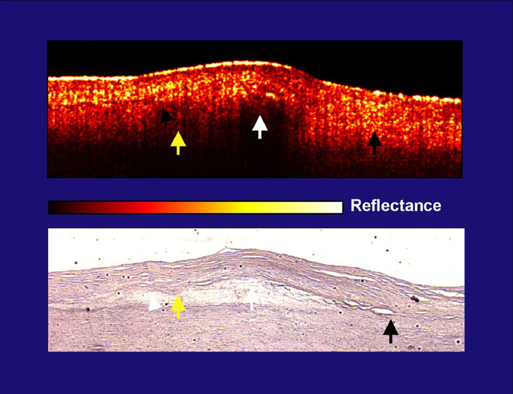
This figure is from the initial Circulation publication from 1996. The yellow arrows demonstrate the cap-lipid interface that is diffuse and is covered by a highly scattering cap (yellow reflections in cap). The white arrow, which is also over lipid, identifies a cap with lower scattering and the cap-lipid interface is sharply defined. Both are over the same lipid collection. Finally, the black arrow shows the intimal-elastic layer interface (no lipid present) that is diffuse, with an intima that is highly scattering. In figure 1B, E, and F of the S-J Kang et. al. paper, there are diffuse border plaques, but the caps over them are highly scattering, which is potentially the etiology. The concern is therefore whether the lipid interface is diffuse in some OCT images because of the core composition, as others have previously suggested, or high scattering from the cap, as we have previously suggested. It is possible that highly scattering caps correlate with lipid plaques, but it is also possible that this scattering is the etiology for the variations in sensitivity and specificity. As clinical studies are now being performed in large numbers due to the availability of the technology, like the paper in this issue, OCT’s lipid characterizing ability needs to be more firmly established.
Comment on
-
Optical coherence tomographic analysis of in-stent neoatherosclerosis after drug-eluting stent implantation.Circulation. 2011 Jun 28;123(25):2954-63. doi: 10.1161/CIRCULATIONAHA.110.988436. Epub 2011 Jun 6. Circulation. 2011. PMID: 21646494
References
-
- Kang SJ, Mintz GS, Akasaka T, et al. Optical Coherent Tomographic Analysis of In-stent Neo-atherosclerosis after Drug-Eluting Stent Implantation. (CIRCULATIONAHA/2010/988436) - PubMed
-
- Brezinski ME. Optical Coherence Tomography: Principle and Practice. Burlington: Academic Press; 2006.
-
- Brezinski ME, Tearney GJ, Bouma BE, Izatt JA, Hee MR, Swanson EA, Southern JF, Fujimoto JG. Optical coherence tomography for optical biopsy - properties and demonstration of vascular pathology. Circulation. 1996;93:1206–1213. - PubMed
-
- St. Jude Medical, St. Paul Minnesota. http://www.sjmprofessional.com /Products/US/Imaging-Diagnostics/OCT-Tech....
-
- Tanigawa J, Barlis P, Dimopoulos K, Dalby M, Moore P, Di Mario C. The influence of strut thickness and cell design on immediate apposition of drug-eluting stents assessed by optical coherence tomography. International Journal Of Cardiology. 2009;134:180–188. - PubMed
Publication types
MeSH terms
Grants and funding
LinkOut - more resources
Full Text Sources
Medical
