. 2011 Aug 1;187(3):1222-34.
doi: 10.4049/jimmunol.1100469. Epub 2011 Jun 27.

Cytosolic DNA-activated human dendritic cells are potent activators of the adaptive immune response

Affiliations

Cytosolic DNA-activated human dendritic cells are potent activators of the adaptive immune response

Katalin Kis-Toth et al. J Immunol. .

Abstract

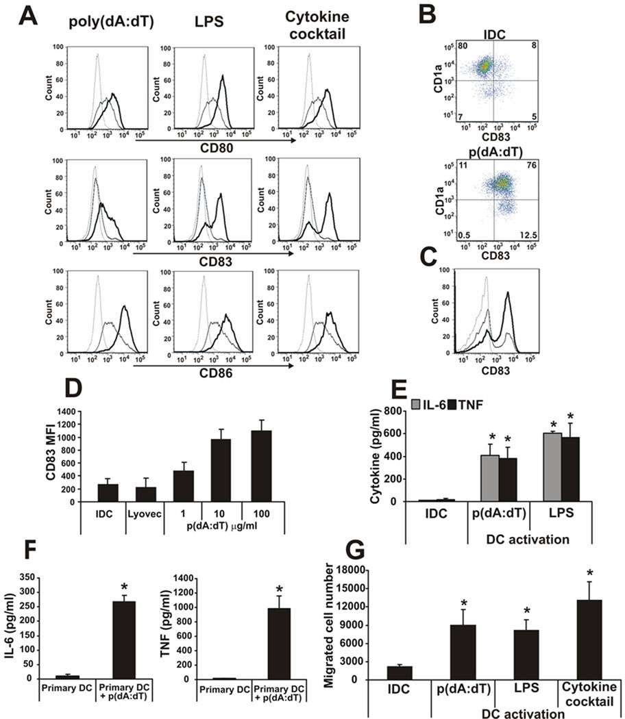
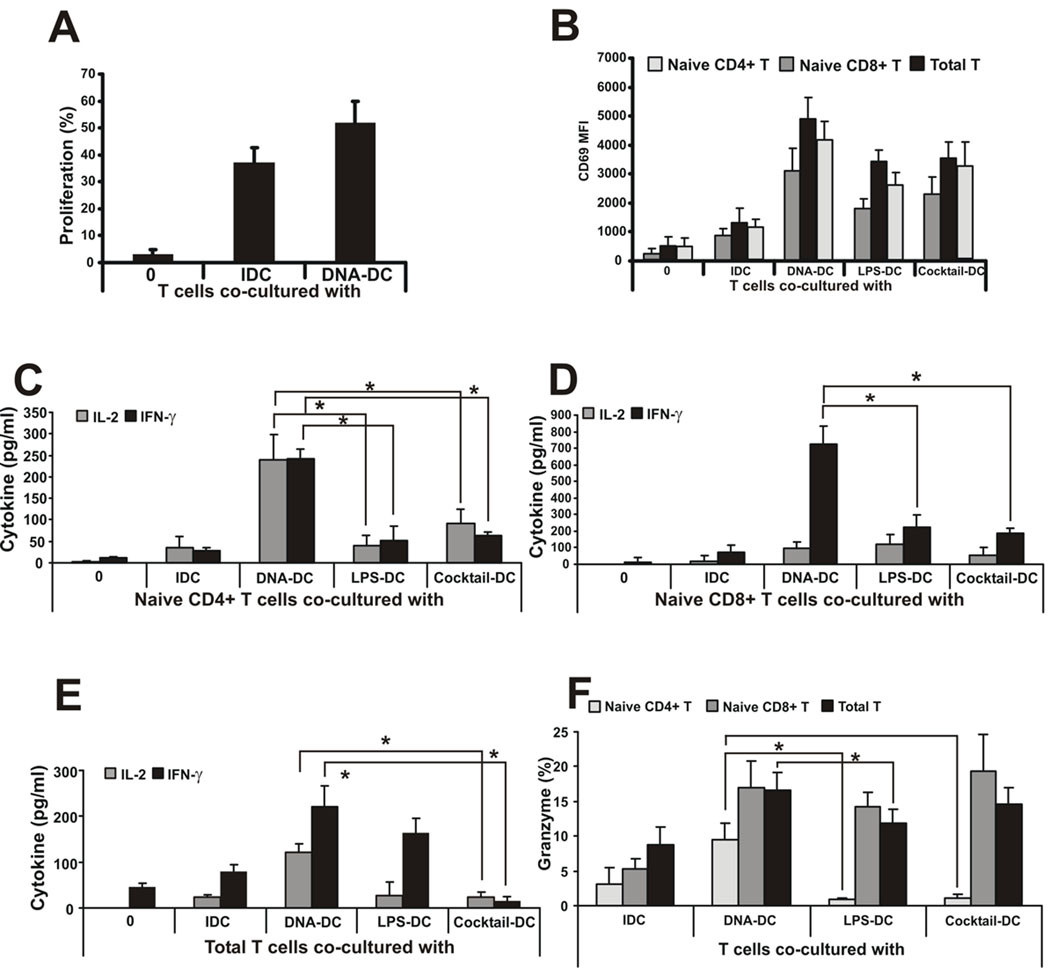
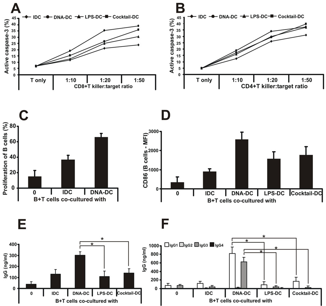
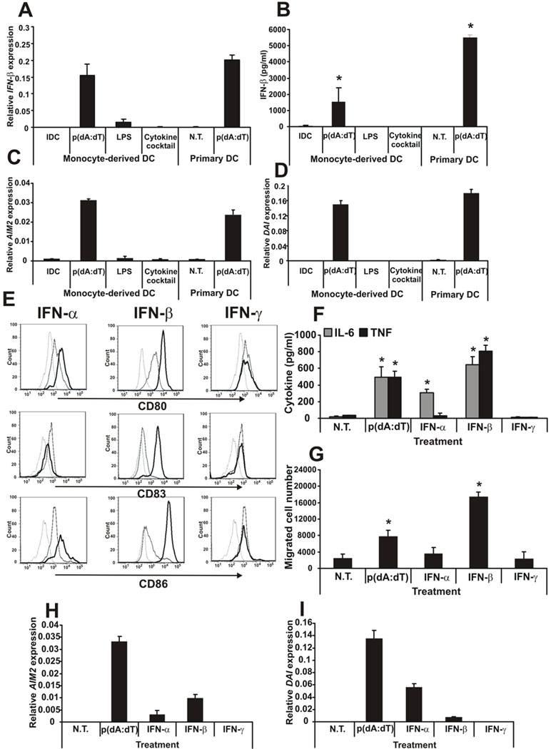
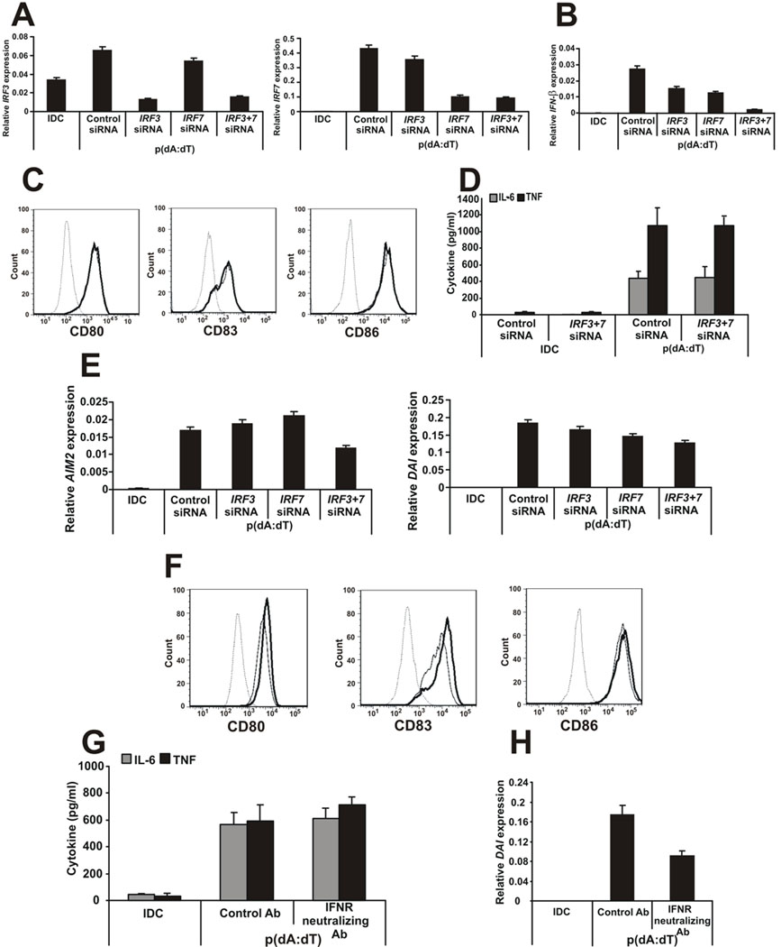
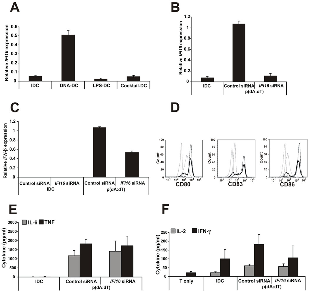
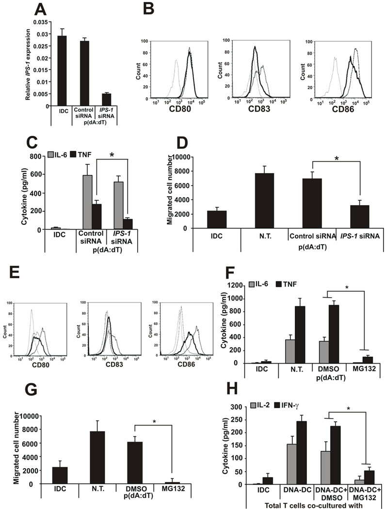
Recent studies in cell lines and genetically engineered mice have demonstrated that cytosolic dsDNA could activate dendritic cells (DCs) to become effector APCs. Recognition of DNA might be a major factor in antimicrobial immune responses against cytosolic pathogens and also in human autoimmune diseases such as systemic lupus erythematosus. However, the role of cytosolic dsDNA in human DC activation and its effects on effector T and B cells are still elusive. In this study, we demonstrate that intracellular dsDNA is a potent activator of human monocyte-derived DCs as well as primary DCs. Activation by dsDNA depends on NF-κB activation, partially on the adaptor molecule IFN-promoter stimulator-1 and the novel cytosolic dsDNA receptor IFI16, but not on the previously recognized dsDNA sentinels absent in melanoma 2, DNA-dependent activator of IFN regulatory factor 3, RNA polymerase III, or high-mobility group boxes. More importantly, we report for the first time, to our knowledge, that human dsDNA-activated DCs, rather than LPS- or inflammatory cytokine mixture-activated DCs, represent the most potent inducers of naive CD4(+) T cells to promote Th1-type cytokine production and generate CD4(+) and CD8(+) cytotoxic T cells. dsDNA-DCs, but not LPS- or mixture-activated DCs, induce B cells to produce complement-fixing IgG1 and IgG3 Abs. We propose that cytosolic dsDNA represents a novel, more effective approach to generate DCs to enhance vaccine effectiveness in reprogramming the adaptive immune system to eradicate infectious agents, autoimmunity, allergy, and cancer.


Figures

Figure 1
Figure 1. Phenotypic characterization of dsDNA-activated DCs
A, Poly(dA:dT) was transfected into monocyte-derived DCs using Lyovec transfection reagent and the level of activations were compared to LPS- and inflammatory cytokine cocktail-activated DCs by CD80, CD83 and CD86 surface expression. Dashed line: isotype control, thin line: immature DC, thick line: activated DC. B, Cytosolic dsDNA activation of CD1a and CD1a+ subpopulations of monocyte-derived DCs and C, primary CD11c+ blood DCs monitored by CD83 upregulation. D, Dose-dependent activation of DCs by poly(dA:dT) monitored by CD83 upregulation. E, IL-6 and TNF pro-inflammatory cytokine production of monocyte-derived DCs and F, primary DCs after poly(dA:dT) transfection. G, Migratory capacity of poly(dA:dT)-, LPS- or inflammatory cytokine cocktail-DCs towards the MDC chemokine MIP3-β. n=5 independent experiments.
Figure 2
Figure 2. dsDNA-activated DCs function as professional APCs
A, Total allogeneic T cells were co-cultured with IDCs or poly(dA:dT)-activated DCs and cell proliferation was measured by CFSE labeling. Proliferation is shown as percentage of cells with low CFSE staining. B, Naïve CD4+, naïve CD8+ and total allogeneic T cells were co-cultured with IDCs or poly(dA:dT)-, LPS- or inflammatory cytokine cocktail-activated DCs and the surface expression of the activation molecule CD69 was measured by flow cytometry. C, Naïve CD4+,D, naïve CD8+ and E, total allogeneic T cells were co-cultured with IDCs or poly(dA:dT)-, LPS- or inflammatory cytokine cocktail-activated DCs and IL-2 and IFN-γ cytokine production by T cells was measured by ELISA. F, Granzyme positive CD4+, CD8+ and total allogeneic T cells co-cultured with differentially activated DCs measured by flow cytometry. N.T.: non-treated. n=4 independent experiments.
Figure 3
Figure 3. dsDNA-activated DCs confer killing ability on CD4+ and CD8+ T cells and collaborate with T cells to generate a specific humoral response
A, Oku-1 cells were co-cultured with naïve CD8+ or B, CD4+ T cells and differentially activated DCs for 4 days at different killer:target ratios (from 1:10 to 1:50). To measure the killing capacity of DC-activated T cells, fresh, CFSE-labeled Oku-1 cells were added to the co-cultures for an additional 4h. To identify apoptotic target cells, flow cytometry was used to detect intracellular active caspase-3 in CFSE-positive cells. n=5 independent experiments. One representative data set is shown. C, Allogeneic B cells were co-cultured with IDCs or poly(dA:dT)-activated DCs and cell proliferation was measured by CFSE labeling. Proliferation is shown as percentage of cells with low CFSE staining. D, Allogeneic B cells were co-cultured with IDCs or poly(dA:dT)-, LPS- or inflammatory cytokine cocktail-activated DCs and the surface expression of CD86 molecule was measured by flow cytometry. E, Allogeneic B and T cells were co-cultured with IDCs or poly(dA:dT)-, LPS- or inflammatory cytokine cocktail-activated DCs and total immunoglobulin G production was measured from the supernatants. F, Immunoglobulin G subclass profiling was done from co-culture supernatants by ELISA. n=3 independent experiments.
Figure 4
Figure 4. dsDNA-activated DCs produce IFN-β and mature upon type I IFN treatment
A, Gene expression and B, protein secretion of IFN-β upon different activation of monocyte-derived and primary DCs. C, AIM2 and D, DAI gene expression upon different activation of monocyte-derived and primary DCs. E, DCs were treated with type I (α and β) and type II (γ) IFNs and the levels of activation were compared by CD80, CD83 and CD86 surface expression. Dashed line: isotype control, thin line: immature DC, thick line: activated DC. F, IL-6 and TNF production and G, migratory capacity of the IFN-activated DCs compared to poly(dA:dT) activation. H, AIM2 and I DAI gene expression in IFN-activated DCs compared to poly(dA:dT) activation. N.T.: non-treated. n=3 independent experiments.
Figure 5
Figure 5. The type I IFN pathway is not required for DC activation by cytosolic dsDNA
A, Effectiveness of IRF3- and IRF7-specific knock-downs. B, IFN-β gene expression in siRNA-treated DNA-DCs. C, CD80, CD83 and CD86 surface expression of the siRNA-treated, DNA-activated DCs. Dashed line: isotype control, thin line: DNA-DC treated with control siRNA, thick line: DNA-DC treated with IRF3- and IRF7-specific siRNAs. D, IL-6 and TNF production by siRNA-treated DNA-DCs. E, AIM2 and DAI gene expression in siRNA-treated DNA-DCs. F, CD80, CD83 and CD86 surface expression of the interferon receptor neutralizing antibody-treated, DNA-activated DCs. Dashed line: isotype control, thin line: DNA-DC treated with control antibody, thick line: DNA-DC treated with interferon receptor-specific neutralizing antibody. G, IL-6 and TNF production of control or neutralizing antibody-treated DNA-DCs. H, DAI gene expression in control or neutralizing antibody-treated DNA-DCs. n=3 independent experiments.
Figure 6
Figure 6. Cytosolic DNA activation of DCs partly depends on the IFI16 sensor
A, IFI16 gene expression upon DC maturation with different activation stimuli. B, Effectiveness of IFI16-specific knock-down. C, IFN-β gene expression by siNA-treated DCs measured after 4h of p(dA:dT) activation by Q-PCR. D, CD80, CD83 and CD86 surface expression of the siRNA-treated, DNA-activated DCs. Dashed line: isotype control, grey thin line: IDC, black thin line: DNA-DC treated with control siRNA, thick line: DNA-DC treated with IFI16-specific siRNA. E, IL-6 and TNF production by siRNA-treated DNA-DCs. F, Total allogeneic T cells were co-cultured with IDCs or siRNA-treated, poly(dA:dT)-activated DCs and IL-2 and IFN-γ cytokine production by T cells was measured by ELISA. n=3 independent experiments.
Figure 7
Figure 7. Cytosolic DNA activation of DCs depends on the IPS-1 and proteosome pathways
A, Effectiveness of IPS-1-specific knock-down. B, CD80, CD83 and CD86 surface expression of the siRNA-treated, DNA-activated DCs. Dashed line: isotype control, thin line: DNA-DC treated with control siRNA, thick line: DNA-DC treated with IPS-1-specific siRNA. C, IL-6 and TNF production by siRNA-treated DNA-DCs. D, Migratory capacity of siRNA-treated DNA-DCs. E, CD80, CD83 and CD86 surface expression of the proteosome inhibitor MG132-treated DCs. Dashed line: isotype control, grey thin line: IDC, black thin line: DNA-DC treated with DMSO, thick line: DNA-DC treated with MG132. F, IL-6 and TNF production and G, migratory capacity of MG132-treated DNA-DCs. H, cytokine production by total T cells co-cultured with MG132-treated DNA-DCs. N.T.: non-treated. n=3 independent experiments.

Similar articles

Cited by

References

    1. Banchereau J, Briere F, Caux C, Davoust J, Lebecque S, Liu YJ, Pulendran B, Palucka K. Immunobiology of dendritic cells. Annu Rev Immunol. 2000;18:767. - PubMed
    1. Lanzavecchia A, Sallusto F. The instructive role of dendritic cells on T cell responses: lineages, plasticity and kinetics. Curr Opin Immunol. 2001;13:291. - PubMed
    1. Kaisho T, Akira S. Critical roles of Toll-like receptors in host defense. Crit Rev Immunol. 2000;20:393. - PubMed
    1. Jefferies CA, Fitzgerald KA. Interferon gene regulation: not all roads lead to Tolls. Trends Mol Med. 2005;11:403. - PubMed
    1. Blasius AL, Beutler B. Intracellular toll-like receptors. Immunity. 2010;32:305. - PubMed

Publication types

MeSH terms