. 2011 Aug;156(4):1808-18.
doi: 10.1104/pp.111.180539. Epub 2011 Jun 27.

The cytokinin receptors of Arabidopsis are located mainly to the endoplasmic reticulum

Affiliations

The cytokinin receptors of Arabidopsis are located mainly to the endoplasmic reticulum

Klaas Wulfetange et al. Plant Physiol. 2011 Aug.

Abstract

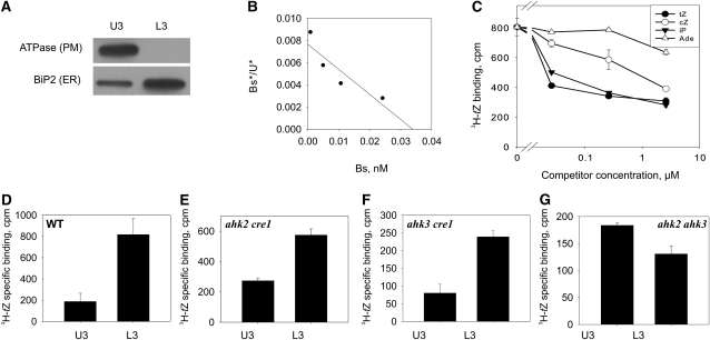
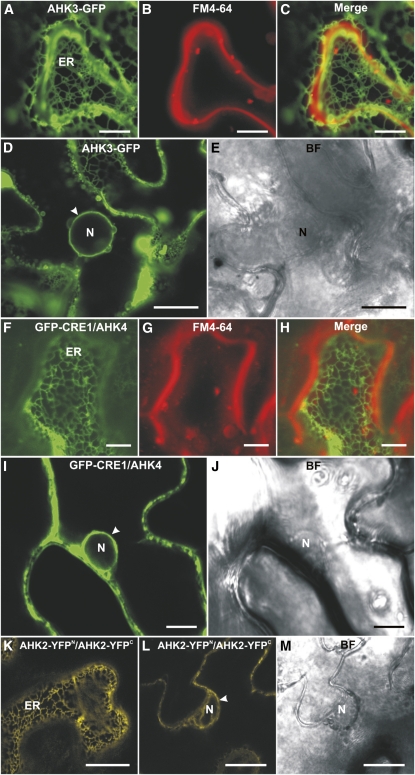
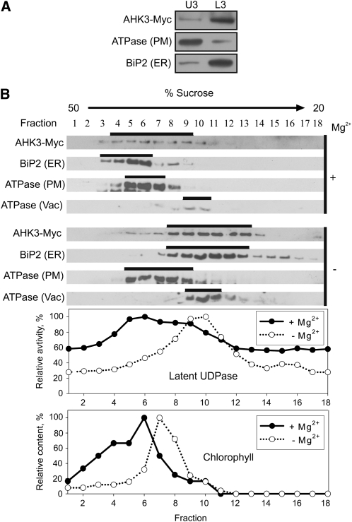
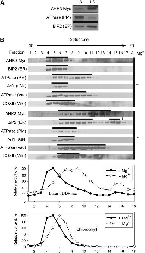
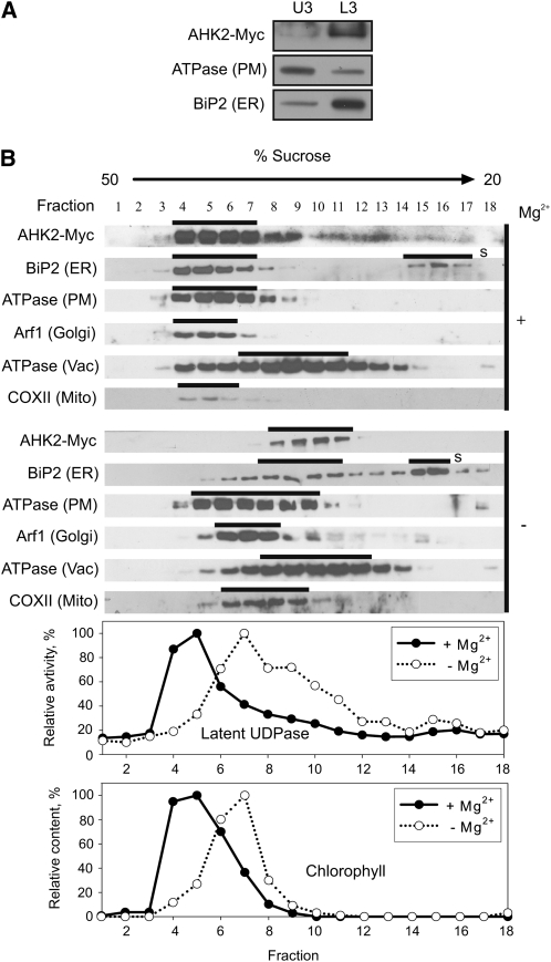
The plant hormone cytokinin is perceived by membrane-located sensor histidine kinases. Arabidopsis (Arabidopsis thaliana) possesses three cytokinin receptors: ARABIDOPSIS HISTIDINE KINASE2 (AHK2), AHK3, and CYTOKININ RESPONSE1/AHK4. The current model predicts perception of the cytokinin signal at the plasma membrane. However, cytokinin-binding studies with membrane fractions separated by two-phase partitioning showed that in the wild type, as well as in mutants retaining only single cytokinin receptors, the major part of specific cytokinin binding was associated with endomembranes. Leaf epidermal cells of tobacco (Nicotiana benthamiana) expressing receptor-green fluorescent protein fusion proteins and bimolecular fluorescence complementation analysis showed strong fluorescence of the endoplasmic reticulum (ER) network for all three receptors. Furthermore, separation of the microsomal fraction of Arabidopsis plants expressing Myc-tagged AHK2 and AHK3 receptors by sucrose gradient centrifugation followed by immunoblotting displayed the Mg²⁺-dependent density shift typical of ER membrane proteins. Cytokinin-binding assays, fluorescent fusion proteins, and biochemical fractionation all showed that the large majority of cytokinin receptors are localized to the ER, suggesting a central role of this compartment in cytokinin signaling. A modified model for cytokinin signaling is proposed.


Figures

Figure 1.
Figure 1.
Cytokinin binding to different membrane fractions obtained by aqueous two-phase partitioning. Six-day-old Arabidopsis seedlings of the wild type and cytokinin receptor double mutants were used for membrane isolation. A, Detection of the membrane-specific markers H+-ATPase (PM) and BiP2 (ER) by immunoblot analysis in the upper (U3) and lower (L3) phases. B, Scatchard plot of [3H]trans-zeatin binding to endomembranes of wild-type plants. Bs*, Specifically bound [3H]trans-zeatin; U*, unbound [3H]trans-zeatin; Bs, total bound trans-zeatin (sum of labeled and unlabeled). The deduced KD is 4.7 nm. C, Competition between [3H]trans-zeatin and unlabeled cytokinins for binding to endomembranes of wild-type plants. Data represent mean values ± se (n = 2). tZ, trans-Zeatin; cZ, cis-zeatin; iP, isopentenyladenine; Ade, adenine. D to G, [3H]trans-Zeatin binding to membrane fractions from the wild type (WT) and double receptor mutants as indicated. U3, Upper phase enriched for PM; L3, lower phase enriched for endomembranes. Data represent mean values ± se (n = 2) of specifically bound [3H]trans-zeatin in cpm per 100 μg of membrane protein.
Figure 2.
Figure 2.
Localization of fluorescent cytokinin receptor fusion proteins in epidermal cells of tobacco leaves. A, ER labeled with AHK3-GFP. B, Staining of the PM with FM4-64 (50 μm). C, Merged image of A and B. D, Localization of AHK3-GFP in the perinuclear space (arrowhead). E, Bright-field (BF) image of D. F, ER labeled with GFP-CRE1/AHK4. G, Staining of the PM with FM4-64 (50 μm). H, Merged image of F and G. I, Localization of GFP-CRE1/AHK4 in the perinuclear space (arrowhead). J, Bright-field image of I. K, BiFC analysis reveals the formation of AHK2-YFPN/AHK2-YFPC homodimers in the ER. L, Homodimerization of AHK2-YFPN and AHK2-YFPC in the perinuclear space (arrowhead). M, Bright-field image of L. N, Nucleus. Bars = 7.5 μm in F to H; 10 μm in A to C, I, and J; and 25 μm in D, E, L, and M.
Figure 3.
Figure 3.
Localization of AHK3-Myc in fractionated membranes from 6-d-old Arabidopsis seedlings expressing PAHK3:AHK3-Myc. A, Aqueous two-phase partitioning of microsomes. Equal amounts of protein from upper (U3) and lower (L3) phases were separated by SDS-PAGE and subjected to immunoblot analysis with antibodies specific for Myc, H+-ATPase (PM), and BiP2 (ER). B, Microsomal membranes were fractionated on linear 20% to 50% (w/w) Suc gradients in the presence of Mg2+ (+) to stabilize ribosomes at the ER or in the absence of Mg2+ (−) to dissociate ribosomes from the ER. Samples (50–100 μL) of each fraction were analyzed by immunoblot using antibodies specific for Myc, H+-ATPase, BiP2, and V-ATPase (Vac, vacuole membrane marker). Latent UDPase activity was used as an enzymatic marker for the Golgi apparatus, and chlorophyll was used to localize thylakoid membranes. In both cases, the highest measured enzyme activity (A650) and chlorophyll content (A652) were set at 100%.
Figure 4.
Figure 4.
Localization of AHK3-Myc in fractionated membranes from shoots of 18-d-old Arabidopsis plants expressing PAHK3:AHK3-Myc. A, Aqueous two-phase partitioning of microsomes. Equal amounts of protein from upper (U3) and lower (L3) phases were separated by SDS-PAGE and subjected to immunoblot analysis with antibodies specific for Myc, H+-ATPase (PM), and BiP2 (ER). B, Microsomal membranes were fractionated on linear 20% to 50% (w/w) Suc gradients in the presence of Mg2+ (+) to stabilize ribosomes at the ER or in the absence of Mg2+ (−) to dissociate ribosomes from the ER. Samples (50–100 μL) of each fraction were analyzed by immunoblot using antibodies specific for Myc, H+-ATPase, BiP2, V-ATPase (Vac, vacuole membrane marker), Arf1 (tGN, trans-Golgi network marker), and COXII (Mito, mitochondria marker). Latent UDPase activity was used as an enzymatic marker for the Golgi apparatus, and chlorophyll was used to localize thylakoid membranes. In both cases, the highest measured enzyme activity (A650) and chlorophyll content (A652) were set at 100%. S, Free soluble BiP2, which has been released during processing and remains at the top of the gradient.
Figure 5.
Figure 5.
Localization of AHK2-Myc in fractionated membranes from Arabidopsis seedlings expressing PAHK2:AHK2-Myc. A, Aqueous two-phase partitioning of microsomes from 6-d-old seedlings. Equal amounts of protein from upper (U3) and lower (L3) phases were separated by SDS-PAGE and subjected to immunoblot analysis with antibodies specific for Myc, H+-ATPase (PM), and BiP2 (ER). B, Microsomal membranes from shoots of 18-d-old plants were fractionated on linear 20% to 50% (w/w) Suc gradients in the presence of Mg2+ (+) to stabilize ribosomes at the ER or in the absence of Mg2+ (−) to dissociate ribosomes from the ER. Samples (50–100 μL) of each fraction were analyzed by immunoblot using antibodies specific for Myc, H+-ATPase, BiP2, V-ATPase (Vac, vacuole membrane marker), Arf1 (Golgi, trans-Golgi network marker), and COXII (Mito, mitochondria marker). Latent UDPase activity was used as an enzymatic marker for the Golgi apparatus, and chlorophyll was used to localize thylakoid membranes. In both cases, the highest measured enzyme activity (A650) and chlorophyll content (A652) were set at 100%. S, Free soluble BiP2, which has been released during processing and remains at the top of the gradient.

References

    1. Argueso CT, Ferreira FJ, Kieber JJ. (2009) Environmental perception avenues: the interaction of cytokinin and environmental response pathways. Plant Cell Environ 32: 1147–1160 - PubMed
    1. Argyros RD, Mathews DE, Chiang YH, Palmer CM, Thibault DM, Etheridge N, Argyros DA, Mason MG, Kieber JJ, Schaller GE. (2008) Type B response regulators of Arabidopsis play key roles in cytokinin signaling and plant development. Plant Cell 20: 2102–2116 - PMC - PubMed
    1. Arnon DI. (1949) Copper enzymes in isolated chloroplasts. Plant Physiol 24: 1–15 - PMC - PubMed
    1. Batoko H, Zheng HQ, Hawes C, Moore I. (2000) A rab1 GTPase is required for transport between the endoplasmic reticulum and Golgi apparatus and for normal Golgi movement in plants. Plant Cell 12: 2201–2218 - PMC - PubMed
    1. Bishopp A, Mähönen AP, Helariutta Y. (2006) Signs of change: hormone receptors that regulate plant development. Development 133: 1857–1869 - PubMed

Publication types

MeSH terms