Review
. 2011 Oct 1;3(10):a005553.
doi: 10.1101/cshperspect.a005553.

Developmental control of oocyte maturation and egg activation in metazoan models

Affiliations
Review

Developmental control of oocyte maturation and egg activation in metazoan models

Jessica R Von Stetina et al. Cold Spring Harb Perspect Biol. .

Abstract

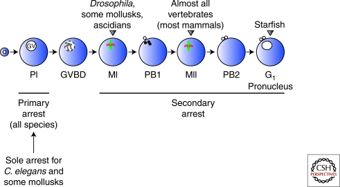
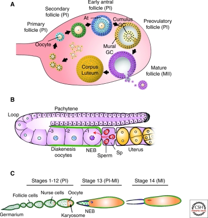
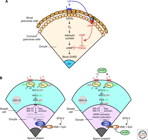
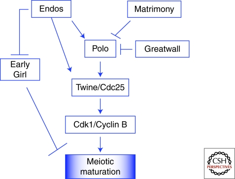
Production of functional eggs requires meiosis to be coordinated with developmental signals. Oocytes arrest in prophase I to permit oocyte differentiation, and in most animals, a second meiotic arrest links completion of meiosis to fertilization. Comparison of oocyte maturation and egg activation between mammals, Caenorhabditis elegans, and Drosophila reveal conserved signaling pathways and regulatory mechanisms as well as unique adaptations for reproductive strategies. Recent studies in mammals and C. elegans show the role of signaling between surrounding somatic cells and the oocyte in maintaining the prophase I arrest and controlling maturation. Proteins that regulate levels of active Cdk1/cyclin B during prophase I arrest have been identified in Drosophila. Protein kinases play crucial roles in the transition from meiosis in the oocyte to mitotic embryonic divisions in C. elegans and Drosophila. Here we will contrast the regulation of key meiotic events in oocytes.


Figures

Figure 1.
Figure 1.
Meiotic arrests during oocyte development. Oocytes from most species undergo a first arrest at prophase I (PI) that is maintained for a few days (Drosophila) or for decades (humans). On hormonal or developmental stimulation, oocytes undergo meiotic maturation, release the primary arrest, and enter a second arrest at metaphase I (MI), metaphase II (MII), or postmeiotic G1 depending on the species. Fertilization triggers release from the second arrest and the completion of meiosis in vertebrates (Xenopus and mammals). Drosophila releases the secondary arrest in a sperm-independent manner. GV, germinal vesicle; PI, prophase I; GVBD, germinal vesicle breakdown; MI, metaphase I; PB1, polar body 1; MII, metaphase II; PB2, polar body 2. (Adapted from Sagata 1996; reprinted with express permission from Noriyuki Sagata.)
Figure 2.
Figure 2.
Stages of oocyte development and meiotic progression. (A) In mammals, a primary follicle, consisting of a prophase I (PI) arrested primary oocyte surrounded by somatic pregranulosa cells, is generated shortly after birth. Primary oocytes grow, whereas granulosa cells (GC) proliferate to form secondary follicles. As a fluid-filled cavity (antrum, At) begins to form, secondary follicles become early antral follicles. In preovulatory follicles, the fully grown primary oocyte is surrounded by cumulus and mural granulosa cells. After a luteinizing hormone surge, the oocyte undergoes meiotic maturation and produces a secondary oocyte that arrests at metaphase II (MII). During ovulation, the MII oocyte is released into the oviduct where on fertilization, meiosis is resumed and completed. (B) In the distal germline of C. elegans, syncytial nuclei enter meiosis and are found in pachytene of PI. Around the loop region, these nuclei cellularize to form oocytes that progress to diakenesis I where they arrest. Somatic-derived gonadal sheath cells (green) surround the developing oocytes. In response to sperm and its secreted factor major sperm protein (MSP), the most proximal oocyte (–1) is induced to undergo meiotic maturation (nuclear envelope breakdown [NEB] and rounding up of the cell). The oocyte passes through the spermatheca (Sp) where fertilization occurs, and it then is deposited into the uterus as a one-cell zygote. (C) The Drosophila oocyte develops within a 16-cell germline cyst surrounded by a monolayer of somatic follicle cells (green). The oocyte enters meiosis in region 2A of the germarium and soon after arrests at PI for most of oogenesis (∼2 d). At stage 13, after a yet unknown developmental or hormonal signal, the oocyte undergoes meiotic resumption and progresses into metaphase I (MI), the secondary arrest point. On ovulation, as the mature stage 14 oocyte travels in the oviduct, rehydration and mechanical pressure trigger the completion of meiosis. (A, Adapted from Matzuk and Lamb 2002; reprinted with express permission from Martin Matzuk. B, Adapted from Kuwabara 2003; reprinted with express permission from Patty Kuwabara. C, Adapted from Xiang et al. 2007; reprinted with express permission from Scott Hawley.)
Figure 3.
Figure 3.
Gap junctional communication between the oocyte and the surrounding somatic cells regulates oocyte meiotic maturation. (A) High levels of cyclic adenosine 3′, 5′-monophosphate (cAMP) in the oocyte inhibit meiotic maturation in mammals. The inhibitory cAMP is produced by the oocyte itself via the activation of the GPR3/Gs/adenylyl cyclase pathway. Guanosine 3′, 5′-cyclic monophosphate (cGMP) produced by the cumulus somatic cells flows through gap junctions into the oocyte to inhibit PDE3A, the phosphodiesterase responsible for the hydrolysis of cAMP. The oocyte stimulates the expression of natriuretic peptide receptor 2 (NPR2) (red), a guanylyl cyclase, in the cumulus cells. Mural granulosa cells induce the generation of cGMP by secreting the NPR2 ligand, natriuretic peptide precursor type C (NPPC) (green). Binding of luteinizing hormone (LH) to its G protein-coupled receptor (GPCR) (blue) reverses the inhibition of meiotic maturation by decreasing the synthesis of cGMP in the somatic follicular layer and by blocking its diffusion through gap junctions. (B) In C. elegans, the Gαo/i pathway in gonadal sheath cells (blue) leads to the inactivation of adenylate cyclase 4 (ACY-4) and subsequently protein kinase A (PKA) in the absence of sperm. This inhibition is postulated to stabilize gonadal sheath-to-oocyte gap junctions composed of innexin (Inx) proteins (Inx-22 and Inx-14), and thereby allow the influx of a negative regulatory signal into the oocyte, which blocks MAP kinase (MAPK) activation and meiotic maturation. In parallel, the VAB-1/Ephrin Receptor in the oocyte inhibits MAPK and oocyte maturation. Sperm-derived major sperm protein (MSP) antagonizes both the sheath Gαo/i and oocyte VAB-1 signaling pathways, while simultaneously activating the sheath Gαs pathway, resulting in MAPK activation and meiotic maturation. It is hypothesized that the Gαs pathway destabilizes sheath-oocyte gap junctions. The nuclear POU homeodomain protein is required for proper differentiation of gonadal sheath cells. (B, Adapted from Govindan et al. 2006 and Sun et al. 2009; reprinted with express permission from Qing-Yuan Sun and David Greenstein, respectively.)
Figure 4.
Figure 4.
Regulatory genes affecting meiotic maturation in Drosophila. Evidence in Drosophila suggests that high levels of Cdk1/Cyclin B activity are required for meiotic maturation. The Polo kinase phosphorylates and activates the phosphatase Twine/Cdc25, which in turn phosphorylates and activates Cdk1. Before nuclear envelope breakdown (NEB), Matrimony sets the timing of meiotic maturation by inhibiting Polo activity. Endos positively regulates the timing of meiotic maturation by regulating the levels of Polo and Twine/Cdc25, which are required to promote Cdk1/Cyclin B activation. Independently, Endos inhibits the predicted E3 ubiquitin ligase Early Girl. The Greatwall kinase also inhibits Polo.

Similar articles

Cited by

References

    1. Abdu U, Brodsky M, Schupbach T 2002. Activation of a meiotic checkpoint during Drosophila oogenesis regulates the translation of Gurken through Chk2/Mnk. Curr Biol 12: 1645–1651 - PubMed
    1. Abrieu A, Brassac T, Galas S, Fisher D, Labbe JC, Doree M 1998. The Polo-like kinase Plx1 is a component of the MPF amplification loop at the G2/M-phase transition of the cell cycle in Xenopus eggs. J Cell Sci 111: 1751–1757 - PubMed
    1. Alphey L, Jimenez J, White-Cooper H, Dawson I, Nurse P, Glover DM 1992. twine, a cdc25 homolog that functions in the male and female germline of Drosophila. Cell 69: 977–988 - PubMed
    1. Archambault V, Zhao X, White-Cooper H, Carpenter AT, Glover DM 2007. Mutations in Drosophila Greatwall/Scant reveal its roles in mitosis and meiosis and interdependence with Polo kinase. PLoS Genet 3: e200. - PMC - PubMed
    1. Avila FW, Ravi Ram K, Bloch Qazi MC, Wolfner MF 2010. Sex peptide is required for the efficient release of stored sperm in mated Drosophila females. Genetics 186: 595–600 - PMC - PubMed

LinkOut - more resources