. 2011 Jul 12;108(28):11590-5.
doi: 10.1073/pnas.1109029108. Epub 2011 Jun 27.

Expression of a functional VEGFR-1 in tumor cells is a major determinant of anti-PlGF antibodies efficacy

Affiliations

Expression of a functional VEGFR-1 in tumor cells is a major determinant of anti-PlGF antibodies efficacy

Jenny Yao et al. Proc Natl Acad Sci U S A. .

Abstract

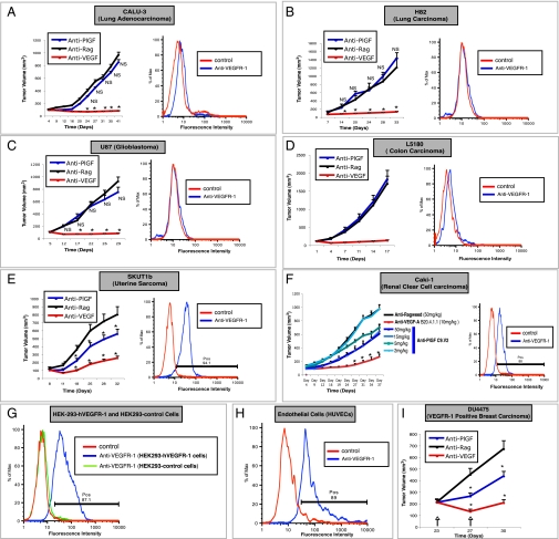
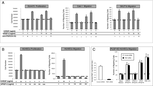
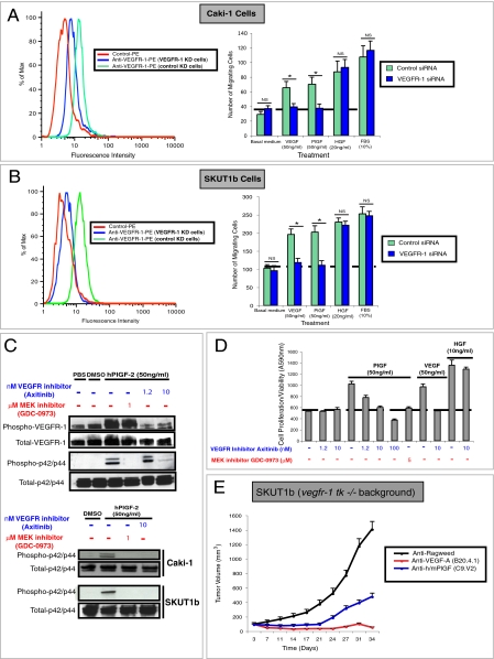
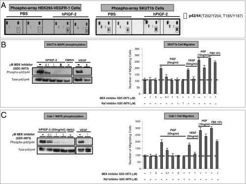
PlGF, one of the ligands for VEGFR-1, has been implicated in tumor angiogenesis. However, more recent studies indicate that genetic or pharmacological inhibition of PlGF signaling does not result in reduction of microvascular density in a variety of tumor models. Here we screened 12 human tumor cell lines and identified 3 that are growth inhibited by anti-PlGF antibodies in vivo. We found that efficacy of anti-PlGF treatment strongly correlates with VEGFR-1 expression in tumor cells, but not with antiangiogenesis. In addition, PlGF induced VEGFR-1 signaling and biological responses in tumor cell lines sensitive to anti-PlGF, but not in refractory tumor cell lines or in endothelial cells. Also, genetic ablation of VEGFR-1 signaling in the host did not affect the efficacy of PlGF blockade. Collectively, these findings suggest that the role of PlGF in tumorigenesis largely consists of promoting autocrine/paracrine growth of tumor cells expressing a functional VEGFR-1 rather than stimulation of angiogenesis.


Conflict of interest statement

The authors declare no conflict of interest.

Figures

Fig. 1.
Fig. 1.
Inhibition of tumor growth by Anti-PlGF mAb treatment is restricted to VEGFR-1 positive xenografts. (AF, Left) Effects of anti-PlGF mAb C9.V2 on primary growth of human tumor xenografts. (F) Dose-dependent inhibition of Caki-1 tumor growth by anti-PlGF C9.V2 Mab. (A–F, Right) Analysis of VEGFR-1 expression in tumor cells. Tumor cells were incubated with biotinylated anti-VEGFR-1 mAb (blue) and or with Streptavidin-PE only as a control (red) as indicated. VEGFR-1 expression was analyzed by flow cytometry. Positive (pos) indicates the calculated percentage of positive cells. (G and H) Flow cytometry VEGFR-1 positive and negative controls. (G) HEK293-VEGFR-1 cells (blue) are VEGFR-1 positive and HEK293-empty vector (green) are VEGFR-1 negative. (H) Endothelial cells (HUVECs) are VEGFR-1 positive (blue). (I) Anti-PlGF inhibits growth of established DU4475 orthotopic breast carcinoma xenografts. Anti-PlGF or anti-Ragweed mAb was given at 15 mg/kg. Anti-VEGF-A mAb was given at 10 mg/kg. All antibody treatments were administrated biweekly. n = 10–15, *P < 0.05 relative to anti-ragweed treatment. Error bars represent SEM.
Fig. 2.
Fig. 2.
Anti-PlGF tumor sensitive tumor-cell lines but not endothelial cells respond to hPlGF-2 stimulation. (A) hPlGF-2 induces dose-dependent biological effects in the anti-PlGF sensitive tumor cell lines DU4475 (Left), CAKI-1 (Center), and SKUT1b (Right), and these effects are blocked by anti-PlGF mAb. (B) hPlGF-2 fails to stimulate HUVEC proliferation (Left) and migration (Right) at all doses tested. (C, Left) Quantification by ELISA of hPlGF released by PlGF knock-down (KD) or control HUVECs. (C, Right) PlGF knock-down (blue bars) and siRNA control HUVECs (green bars) remain unresponsive to hPlGF-2. The figure shows the average values from representative experiments. Doses of ligands are indicated in the figure. Dotted lines represent basal (control) activity. Experiments were repeated at least three times with comparable results. n = 3–5. Error bars represent SD.
Fig. 4.
Fig. 4.
Inhibition of PlGF/VEGFR-1 signaling in tumor but not stromal cells is a major determinant for anti-PlGF efficacy. (A and B, Left) VEGFR-1 siRNA but not control siRNA reduces VEGFR-1 expression by FACS. (A and B, Right) Effects of VEGFR-1 knock-down on migration of Caki-1 and SKUT1b cells in response to PlGF, VEGF, HGF, or 10% FBS. (C, Upper) Effects of axitinib (VEGFR inhibitor) and GDC-0973 on hPlGF-2-induced phosphorylation of VEGFR-1 and p42/p44 in HEK293-VEGFR-1 cells. (C, Lower) Effects of axinitinib and GDC-0973 on hPlGF-2-induced phosphorylation of p42/p44 in HEK-293-CAKI-1 and SKUT1b cells. (D) Effects of axinitinib and GDC-0973 on hPlGF-2-induced proliferation/survival of SKUT1b cells. Dotted lines represent basal levels of migration or proliferation. Experiments were repeated at least three times with comparable results. n = 3–5. Error bars represent SD. (E) Effects of anti-PlGF, anti-VEGF-A, or anti-ragweed mAb on the growth of tumors implanted in vegfr-1 tk−/−, rag2−/− mice. Antibodies were administered as indicated in Fig. 1 and in Materials and Methods. n = 10, relative to anti-ragweed treatment. Error bars represent SEM.
Fig. 3.
Fig. 3.
hPlGF-2-induced responses in anti-PlGF sensitive cell lines require MAPK activation. (A) Phospho-antibody array analyses of hPlGF or mock-stimulated HEK293-VEGFR-1 (Left) and SKUT1b (Right) cells. The figure shows only a relevant section of phopho-array membrane. (B, Left) Effects of MEK inhibitor GDC-0973 on PlGF-induced MAPK phosphorylation in SKUT1b cells. (B, Right) Effects of GDC-0973 or RAF inhibitor (GDC-0879) on PlGF-induced SKUT1b cell migration. (C, Left) Effect of MEK inhibitor on PlGF-induced MAPK phosphorylation in CAKI-1 cells. (C, Right) Effects of MEK inhibitor and RAF inhibitor on PlGF-induced Caki-1 cell migration. Dotted lines represent basal (control) activity. Experiments were repeated at least three times with comparable results. n = 3–5. Error bars represent SD.

References

    1. Ferrara N, Gerber HP, LeCouter J. The biology of VEGF and its receptors. Nat Med. 2003;9:669–676. - PubMed
    1. Fischer C, Mazzone M, Jonckx B, Carmeliet P. FLT1 and its ligands VEGFB and PlGF: drug targets for anti-angiogenic therapy? Nat Rev Cancer. 2008;8:942–956. - PubMed
    1. Lichtenberger BM, et al. Autocrine VEGF signaling synergizes with EGFR in tumor cells to promote epithelial cancer development. Cell. 2010;140:268–279. - PubMed
    1. Wu Y, et al. The vascular endothelial growth factor receptor (VEGFR-1) supports growth and survival of human breast carcinoma. Int J Cancer. 2006;119:1519–1529. - PubMed
    1. Sawano A, Takahashi T, Yamaguchi S, Aonuma M, Shibuya M. Flt-1 but not KDR/Flk-1 tyrosine kinase is a receptor for placenta growth factor, which is related to vascular endothelial growth factor. Cell Growth Differ. 1996;7:213–221. - PubMed

MeSH terms

LinkOut - more resources