Case Reports
. 2011 Jul 12;108(28):11638-43.
doi: 10.1073/pnas.1109144108. Epub 2011 Jun 27.

Resting-state activity in development and maintenance of normal brain function

Affiliations
Case Reports

Resting-state activity in development and maintenance of normal brain function

Carolyn E Pizoli et al. Proc Natl Acad Sci U S A. .

Abstract

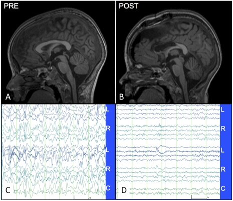
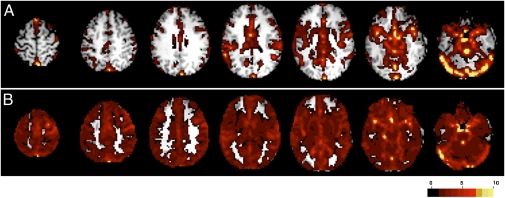
One of the most intriguing recent discoveries concerning brain function is that intrinsic neuronal activity manifests as spontaneous fluctuations of the blood oxygen level-dependent (BOLD) functional MRI signal. These BOLD fluctuations exhibit temporal synchrony within widely distributed brain regions known as resting-state networks. Resting-state networks are present in the waking state, during sleep, and under general anesthesia, suggesting that spontaneous neuronal activity plays a fundamental role in brain function. Despite its ubiquitous presence, the physiological role of correlated, spontaneous neuronal activity remains poorly understood. One hypothesis is that this activity is critical for the development of synaptic connections and maintenance of synaptic homeostasis. We had a unique opportunity to test this hypothesis in a 5-y-old boy with severe epileptic encephalopathy. The child developed marked neurologic dysfunction in association with a seizure disorder, resulting in a 1-y period of behavioral regression and progressive loss of developmental milestones. His EEG showed a markedly abnormal pattern of high-amplitude, disorganized slow activity with frequent generalized and multifocal epileptiform discharges. Resting-state functional connectivity MRI showed reduced BOLD fluctuations and a pervasive lack of normal connectivity. The child underwent successful corpus callosotomy surgery for treatment of drop seizures. Postoperatively, the patient's behavior returned to baseline, and he resumed development of new skills. The waking EEG revealed a normal background, and functional connectivity MRI demonstrated restoration of functional connectivity architecture. These results provide evidence that intrinsic, coherent neuronal signaling may be essential to the development and maintenance of the brain's functional organization.


Conflict of interest statement

The authors declare no conflict of interest.

Figures

Fig. 1.
Fig. 1.
Structural MRI (midline sagittal T1-weighted MP-RAGE) and EEG. (A) Preoperative MRI shows normal structure. (B) Postoperative MRI (on postoperative day 1) shows the extent of the anterior two-thirds corpus callosotomy. Widening of the interhemispheric fissure, a common postoperative finding, also is evident. (C) Waking EEG (10 μV/mm, 1 s spacing) recorded 6 mo preoperatively using a standard (“double banana”) bipolar montage. The record is severely abnormal (see text). (D) Normal waking EEG recorded 4 mo postoperatively. “L,” “R,” and “C” denote left, right, and central derivations. The montage, time, and amplitude scales are identical in C and D.
Fig. 2.
Fig. 2.
Selected seed-based correlation maps. Columns show the seeds (Left), preoperative maps (Middle), and postoperative maps (Right). The map quantity illustrated is the Fisher z-transformed correlation coefficient thresholded at ± 0.2. (A) Left somatomotor cortex seed (−39 −26 51); somatomotor RSN. (B) Left posterior cingulate/precuneus seed (−4 −40 43); default mode network (DMN). (C) Visual cortex seed (−20 −75 12). (D) Auditory cortex seed (−50 −25 8). (E) Left inferior frontal gyrus seed (−48 −13 31); speech. (F) Left intraparietal sulcus seed (−24 −69 30); dorsal attention network. Note the marked improvement in RSN organization in the postoperative maps vs. the preoperative maps.
Fig. 3.
Fig. 3.
Voxel-wise SD maps. (A) Preoperative results. High signal variation is seen primarily in vascular and CSF spaces. (B) Postoperative results. High signal variation is seen primarily in gray matter.
Fig. 4.
Fig. 4.
Correlation and covariance matrices corresponding to 46 ROIs taken pairwise. The ROIs are ordered according to functional networks delineated by brackets and depicted on the right; see SI Text for details. (A) Preoperative correlation. (B) Postoperative correlation. (C) Preoperative covariance. (D) Postoperative covariance. Note the improved RSN organization evident in the block structure of the postoperative results, especially comparing C and D. The diagonals in D and B show a pronounced postoperative increase in BOLD signal variance.

Similar articles

Cited by

References

    1. Purdon PL, Weisskoff RM. Effect of temporal autocorrelation due to physiological noise and stimulus paradigm on voxel-level false-positive rates in fMRI. Hum Brain Mapp. 1998;6:239–249. - PMC - PubMed
    1. Biswal B, Yetkin FZ, Haughton VM, Hyde JS. Functional connectivity in the motor cortex of resting human brain using echo-planar MRI. Magn Reson Med. 1995;34:537–541. - PubMed
    1. Smith SM, et al. Correspondence of the brain's functional architecture during activation and rest. Proc Natl Acad Sci USA. 2009;106:13040–13045. - PMC - PubMed
    1. Cole DM, Smith SM, Beckmann CF. Advances and pitfalls in the analysis and interpretation of resting-state FMRI data. Front Syst Neurosci. 2010;4:8. - PMC - PubMed
    1. Biswal BB, et al. Toward discovery science of human brain function. Proc Natl Acad Sci USA. 2010;107:4734–4739. - PMC - PubMed

Publication types

MeSH terms

Supplementary concepts