. 2011 Jul 12;108(28):11458-63.
doi: 10.1073/pnas.1017474108. Epub 2011 Jun 27.

Myosin-II inhibition and soft 2D matrix maximize multinucleation and cellular projections typical of platelet-producing megakaryocytes

Affiliations

Myosin-II inhibition and soft 2D matrix maximize multinucleation and cellular projections typical of platelet-producing megakaryocytes

Jae-Won Shin et al. Proc Natl Acad Sci U S A. .

Abstract

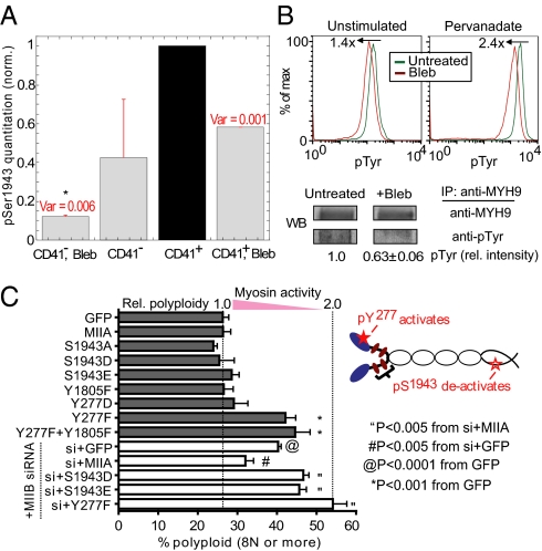
Cell division, membrane rigidity, and strong adhesion to a rigid matrix are all promoted by myosin-II, and so multinucleated cells with distended membranes--typical of megakaryocytes (MKs)--seem predictable for low myosin activity in cells on soft matrices. Paradoxically, myosin mutations lead to defects in MKs and platelets. Here, reversible inhibition of myosin-II is sustained over several cell cycles to produce 3- to 10-fold increases in polyploid MK and a number of other cell types. Even brief inhibition generates highly distensible, proplatelet-like projections that fragment readily under shear, as seen in platelet generation from MKs in vivo. The effects are maximized with collagenous matrices that are soft and 2D, like the perivascular niches in marrow rather than 3D or rigid, like bone. Although multinucleation of other primary hematopoietic lineages helps to generalize a failure-to-fission mechanism, lineage-specific signaling with increased polyploidy proves possible and novel with phospho-regulation of myosin-II heavy chain. Label-free mass spectrometry quantitation of the MK proteome uses a unique proportional peak fingerprint (ProPF) analysis to also show upregulation of the cytoskeletal and adhesion machinery critical to platelet function. Myosin-inhibited MKs generate more platelets in vitro and also in vivo from the marrows of xenografted mice, while agonist stimulation activates platelet spreading and integrin αIIbβ3. Myosin-II thus seems a central, matrix-regulated node for MK-poiesis and platelet generation.


Conflict of interest statement

The authors declare no conflict of interest.

Figures

Fig. 1.
Fig. 1.
Myosin affects MK maturation and cell fragmentation. (A) In vivo scheme of MK-poiesis and platelet fragmentation in bone marrow. (B) Modeling MK maturation and platelet fragmentation in vitro by myosin inhibition and micropipette aspiration on CD34+-derived cultures. (C) Myosin inhibition by blebbistatin accelerates CD41+ MK polyploidization. Representative flow cytometry plots for ploidy analysis (Upper), and dose-dependence (Lower). Absolute values were normalized to 104 initial cell input (n ≥ 4 donors, ± SEM). (D) Myosin inhibition increases membrane extension and fragmentation in micropipette aspiration. Representative fragmentation within seconds (Upper) after 30 min of 20 μM blebbistatin treatment and aspiration ΔP = 1.4 kPa. Aspiration length vs. effective cortical tension (Lower) with median results shown (Fig. S2A).
Fig. 2.
Fig. 2.
Effects of matrix elasticity and ligand density on MK polyploidy. All MK polyploid cell numbers were scaled to 104 initial cell input (n ≥ 3 donors; ± SEM). (A) Soft (0.3 kPa) matrices always facilitate MK polyploidization on 2 ng/cm2 (low) collagen gels. Tukey’s HSD Test indicates P < 0.05 for all pairs except 0.3 kpa 0 μM vs. 34 kpa 20 μM. (B) On a range of collagen concentrations, ratios of polyploid MK numbers for soft (0.3 kPa) versus stiff (34 kPa) matrices fit to standard dose-response curves: untreated and blebbistatin-treated IC50 ∼20, 1,000 ng/cm2, respectively (Hill coefficients ∼ −1.5, −2.0 respectively). (C) Cell adhesion on soft and stiff matrices after 3 d cell culture ± blebbistatin. *P < 0.05 for 0.3 kPa vs. 34 kPa untreated. (D) Polyploid MK numbers on stiff gels cultured on a range of collagen concentrations. Dose-response curves are shown for untreated (EC50 ∼200 ng/cm2, Hill coefficient = 1.0) and blebbistatin-treated cells (IC50 ∼0.3 ng/cm2, Hill coefficient = −1.3). Cartoon depicts effects on cell adhesion and division. (E) Proplatelet formation with different gels. (Left) Representative images with F-actin (red). (Scale bar, 5 μm.) (Right) Quantitation of branch length from cell body. (>50 measurements for each group, n ≥ 2 donors). 3D matrices are soft with E ∼1 to 3 kPa. P values are reported from Tukey’s HSD test.
Fig. 3.
Fig. 3.
Sustained inhibition of myosin-II blocks cytokinesis. (A) Generation of polyploid cells is exponential in duration of exposure to 20 μM blebbistatin with doubling time of 18.2 h. All values were scaled to an initial cell input of 104 cells (n = 3, ± SEM). (B) Live cell imaging shows reversal of cytokinesis with blebbistatin for ∼80% of cells observed; without drug, all cells divided. (Scale bar, 10 μm.)
Fig. 4.
Fig. 4.
Phosphorylation of NMM-IIA regulates polyploidization. (A) MS analyses of phospho-S1943 in primary cells. Each pS1943 signal was normalized first to total NMM-IIA signal, which was then normalized to values from CD41+ sorted cells (untreated), and averaged between experiments (n = 2). Ion current of pS1943 in CD41+ is ∼1% of total NMM-IIA signal. *P < 0.05 from one-way ANOVA with Tukey’s HSD test. (B) Blebbisatin treatment leads to overall reduction of pTyr levels under both unstimulated and pervanadate (100 μM)-stimulated conditions as assessed by flow cytometry (Upper). IP of NMM-IIA from lysates of cells treated with pervanadate ± blebbistatin, followed by immunoblot of pTyr and densitometry (Lower) shows reduced pTyr levels in NMM-IIA head (150 kDa) region (n = 3, ± SEM). (C) NMM-IIA head pTyr mutant (Y277F) increases polyploidization. COS cells were transfected as indicated ± pretransfection with NMM-IIB siRNA. Cultured cells were stained with Hoechst 33342 to quantify polyploidy (≥8 N) by flow cytometry (n ≥ 3, ± SEM).
Fig. 5.
Fig. 5.
Label-free MS quantitation of myosin-inhibited cytoskeletal proteome. CD34+ cells were treated with blebbistatin for 3 d. The viable cell fraction (Annexin-V and 7-AAD) of CD41+ MKs was isolated by sorting, followed by MS (see SI Materials and Methods, Fig. S7B, and Dataset S1D). The first column summarizes detectability in prior literature on platelets. <PRF> refers to the number of peptides retained in propotional peak fingerprint for protein quantitation, whereas “Total” refers to all peptides detected.
Fig. 6.
Fig. 6.
Platelets derived from myosin-inhibited MKs. (A) Platelets per CD41+ cell. (Left) NSG mice transplanted intratibially with human-CD34+-derived cells that were pretreated ex vivo with blebbistatin for 3 d (versus untreated cells) show enhanced circulating human platelets. (P < 0.0001; n = 9 mice ± SEM). (Right) MKs exposed to blebbistatin (20 μM for 3 d, then 3 d of no drug) show more in vitro platelets (P < 0.05; n = 5 donors, ± SEM). (B) Similar immunostaining of microtubules in the various MK-derived platelets (n = 3 donors). (C) Similar adhesion and filipodia formation of various MK-derived platelets on collagen-I upon thrombin (1 U/mL) stimulation. (n = 3 donors). (D) Platelets derived from blebbistatin-exposed MKs show normal PAC-1 binding with thrombin (1 U/mL). Tirofiban (10 μM) selectively antagonizes αIIbβ3 and inhibits as expected (35). Representative flow cytometry plots from three experiments. (Scale bars, 5 μm.)

Similar articles

Cited by

References

    1. Straight AF, et al. Dissecting temporal and spatial control of cytokinesis with a myosin II Inhibitor. Science. 2003;299:1743–1747. - PubMed
    1. Engler AJ, Sen S, Sweeney HL, Discher DE. Matrix elasticity directs stem cell lineage specification. Cell. 2006;126:677–689. - PubMed
    1. Conti MA, Even-Ram S, Liu C, Yamada KM, Adelstein RS. Defects in cell adhesion and the visceral endoderm following ablation of nonmuscle myosin heavy chain II-A in mice. J Biol Chem. 2004;279:41263–41266. - PubMed
    1. Zang JH, et al. On the role of myosin-II in cytokinesis: Division of Dictyostelium cells under adhesive and nonadhesive conditions. Mol Biol Cell. 1997;8:2617–2629. - PMC - PubMed
    1. De Lozanne A, Spudich JA. Disruption of the Dictyostelium myosin heavy chain gene by homologous recombination. Science. 1987;236:1086–1091. - PubMed

Publication types

MeSH terms

LinkOut - more resources