. 2011 Jul 12;108(28):11476-81.
doi: 10.1073/pnas.1104161108. Epub 2011 Jun 27.

Myosin VIIa and sans localization at stereocilia upper tip-link density implicates these Usher syndrome proteins in mechanotransduction

Affiliations

Myosin VIIa and sans localization at stereocilia upper tip-link density implicates these Usher syndrome proteins in mechanotransduction

M'hamed Grati et al. Proc Natl Acad Sci U S A. .

Abstract

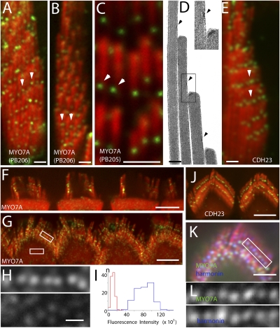
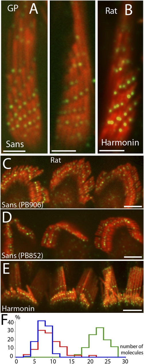
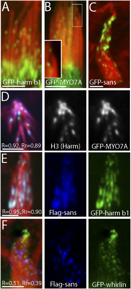
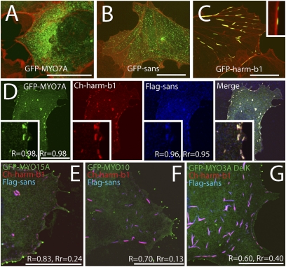
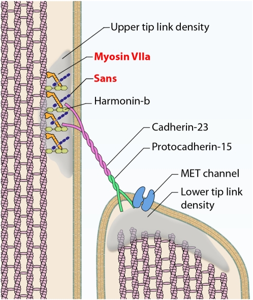
In the most accepted model for hair cell mechanotransduction, a cluster of myosin motors located at the stereocilia upper tip-link density (UTLD) keeps the tip-link under tension at rest. Both myosin VIIa (MYO7A) and myosin 1c have been implicated in mechanotransduction based on functional studies. However, localization studies are conflicting, leaving open the question of which myosin localizes at the UTLD and generates the tip-link resting tension. Using immunofluorescence, we now show that MYO7A and sans, a MYO7A-interacting protein, cluster at the UTLD. Analysis of the immunofluorescence intensity indicates that eight or more MYO7A molecules are present at each UTLD, consistent with a direct role for MYO7A in maintaining tip-link tension. MYO7A and sans localization at the UTLD is confirmed by transfection of hair cells with GFP-tagged constructs for these proteins. Cotransfection studies in a heterologous system show that MYO7A, sans, and the UTLD protein harmonin-b form a tripartite complex and that each protein is capable of interacting with one another independently. We propose that MYO7A, sans, and harmonin-b form the core components of the UTLD molecular complex. In this complex, MYO7A is likely the motor element that pulls on CDH23 to exert tension on the tip-link.


Conflict of interest statement

The authors declare no conflict of interest.

Figures

Fig. 1.
Fig. 1.
Localization of MYO7A to the UTLD. (AC) Immunofluorescence confocal images of MYO7A using antibodies PB206 and PB205 in vestibular hair cells of guinea pig showing labeling (green, arrowheads) at the UTLD location. Stereocilia are counterstained in red with rhodamine phalloidin. (D) UTLD (arrowheads and Inset) in a thin section electron micrograph of a vestibular stereocilia bundle. (E) CDH23 labeling in vestibular hair cells is similar to that seen for MYO7A. (F and G) MYO7A and (J) CDH23 labeling at the UTLD in rat outer hair cells. (H) Close-up views of the areas indicated by the rectangles show MYO7A fluorescence puncta at the UTLD (Upper) and in the cytoplasm (Lower). (I) Distribution of the MYO7A fluorescence intensity at the UTLD (blue) and cytoplasm (red) puncta. (K) Coimmunofluorescence labeling of MYO7A (green) using the sc-74516 antibody and harmonin (blue) in rat outer hair cell. (L) Close-up view of green channel (Upper) and the blue channel (Lower) of the rectangle in K show colocalization of the two proteins. [Scale bars: 1 μm (A–C, E, and K), 5 μm (F, G, and J), and 300 nm (D and H).]
Fig. 2.
Fig. 2.
Localization of sans at the UTLD. (A) Immunofluorescence of sans in guinea pig vestibular hair cells using antibody PB852 shows localization (green puncta) corresponding to UTLD location. (B) Immunofluorescence of harmonin (green) in rat vestibular hair cells shows localization at the UTLD. (C and D) Immunofluorescence of sans in rat outer hair cells using antibodies PB906 (C) and PB852 (D) shows localization (green) in two rows across the tallest and middle stereocilia. (E) Harmonin labeling in rat outer hair cells. (F) Distribution of the estimated number of MYO7A (blue) sans (red) and harmonin (green) at the UTLD based on the ratio of fluorescence intensity at the UTLD puncta and the intensity of the MYO7A cytoplasmic puncta assuming that it represents the fluorescence intensity that corresponds to a single epitope in our assays (n = 100). [Scale bars: 2 μm (A and B) and 5 μm (C–E).]
Fig. 3.
Fig. 3.
Harmonin, MYO7A, and sans form clusters and colocalize in plaques along the stereocilia of transfected vestibular hair cells. GFP-harmonin-b (A), GFP-MYO7A (B), and GFP-sans (C) appear in plaques (green) along the side of stereocilia (counterstained in red with rhodamine phalloidin). (D) Hair cell expressing GFP-MYO7A (green) and counterstained for harmonin with H3 antibody (blue) show colocalization (R = 0.92, Rr = 0.89) of these proteins. (E) Hair cell coexpressing GFP-harmonin-b (green) and Flag-tagged sans (blue) also shows colocalization (R = 0.95, Rr = 0.90) in plaques along the stereocilia. (F) Hair cell coexpressing GFP-whirlin (green) and Flag-tagged sans (blue) shows that GFP-whirlin localizes to the stereocilia tips and does not overlap (R = 0.51, Rr = 0.39) with sans localization presumably at the UTLD. (Scale bars: 2 μm.)
Fig. 4.
Fig. 4.
Tripartite interactions of MYO7A, sans and harmonin in COS7 cells. (A) GFP-MYO7A (green) shows a diffuse cytoplasmic distribution with no obvious colocalization with actin (counterstained in red with rhodamine phalloidin). (B) In COS7 cells, GFP-sans (green) shows granular distribution in the cytoplasm unrelated to actin bundles (red). (C) GFP-harmonin-b (green) form plaques associated with actin (red). (Inset) Close-up view. (D) COS7 cells expressing GFP-MYO7A (green), Cherry-harmonin-b (red), and Flag-sans (blue) show colocalization of MYO7A and sans in the actin-associated harmonin-b plaques (R = 0.98, Rr = 0.98 and R = 0.96, Rr = 0.95, respectively). COS7 cells expressing Cherry-harmonin-b (red) and Flag-sans (blue) together with GFP-MYO15A (E), GFP-MYO10 (F), or GFP-MYO3A (G) show low Pearson’s coefficients (Rr) values indicative of weak and random colocalization. Neither harmonin nor sans colocalize with GFP-MYO15A (E), GFP-MYO10 (F) or GFP-MYO3A (G) at filopodia tips. (Scale bars: 5 μm.)
Fig. 5.
Fig. 5.
Diagram of the mechanotransduction complex illustrating the localization of MYO7A and sans at the upper tip-link density. MYO7A localization is consistent with a role in tip-link tensing.

References

    1. Grillet N, et al. The mechanotransduction machinery of hair cells. Sci Signal. 2009;2:pt5. - PubMed
    1. Gillespie PG, Müller U. Mechanotransduction by hair cells: Models, molecules, and mechanisms. Cell. 2009;139:33–44. - PMC - PubMed
    1. Sakaguchi H, Tokita J, Müller U, Kachar B. Tip links in hair cells: Molecular composition and role in hearing loss. Curr Opin Otolaryngol Head Neck Surg. 2009;17:388–393. - PMC - PubMed
    1. Kazmierczak P, et al. Cadherin 23 and protocadherin 15 interact to form tip-link filaments in sensory hair cells. Nature. 2007;449:87–91. - PubMed
    1. Kachar B, Parakkal M, Kurc M, Zhao Y, Gillespie PG. High-resolution structure of hair-cell tip links. Proc Natl Acad Sci USA. 2000;97:13336–13341. - PMC - PubMed

Publication types

MeSH terms