. 2011 Jul 12;108(28):11668-73.
doi: 10.1073/pnas.1102984108. Epub 2011 Jun 27.

Adjacent visual representations of self-motion in different reference frames

Affiliations

Adjacent visual representations of self-motion in different reference frames

David Mattijs Arnoldussen et al. Proc Natl Acad Sci U S A. .

Abstract

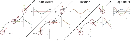
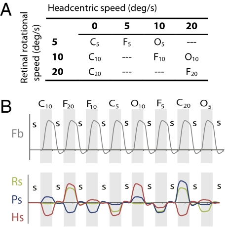
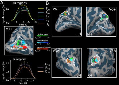
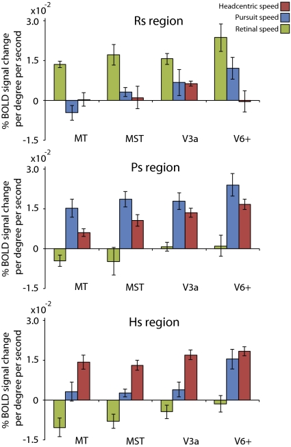
Recent investigations indicate that retinal motion is not directly available for perception when moving around [Souman JL, et al. (2010) J Vis 10:14], possibly pointing to suppression of retinal speed sensitivity in motion areas. Here, we investigated the distribution of retinocentric and head-centric representations of self-rotation in human lower-tier visual motion areas. Functional MRI responses were measured to a set of visual self-motion stimuli with different levels of simulated gaze and simulated head rotation. A parametric generalized linear model analysis of the blood oxygen level-dependent responses revealed subregions of accessory V3 area, V6(+) area, middle temporal area, and medial superior temporal area that were specifically modulated by the speed of the rotational flow relative to the eye and head. Pursuit signals, which link the two reference frames, were also identified in these areas. To our knowledge, these results are the first demonstration of multiple visual representations of self-motion in these areas. The existence of such adjacent representations points to early transformations of the reference frame for visual self-motion signals and a topography by visual reference frame in lower-order motion-sensitive areas. This suggests that visual decisions for action and perception may take into account retinal and head-centric motion signals according to task requirements.


Conflict of interest statement

The authors declare no conflict of interest.

Figures

Fig. 1.
Fig. 1.
Visualization of the stimuli for the three different conditions together with plots of the gaze (green), eye-in-head (blue), and head-in-space (red) orientation at different instants in time. The flow field simulates self-motion through space in a forward direction (the global heading direction, black arrow), but along an oscillating path (black trajectory). Rotation of the eyes and head are defined relative to the global heading direction (ω). The gaze rotation is identical in the three conditions. At every time point, gaze is aligned with the tangent of the path (green arrow). As a result, the flow patterns on the retina remain the same. Combined with different eye-in-head positions, however, different simulated head rotations (red arrows) are defined. At the three instances depicted, the direction of motion along the path deviates maximally from the global heading direction. In the CONSISTENT condition, the amplitude and direction of simulated gaze rotation matches the amplitude and direction of the eye-in-head rotation. Thus, the simulated head rotation is zero. In the FIXATION condition, eye and head remain aligned with the momentary heading direction. Therefore, simulated gaze and head rotation are identical. In the OPPONENT condition, simulated gaze rotation is always opposite in direction to the eye-in-head rotation. Now, the eye rotation relative to the global heading direction remains the same, but the magnitude of the simulated head rotation is doubled. Note that the angles of rotation are exaggerated for visualization purposes.
Fig. 2.
Fig. 2.
Stimulus conditions in the main experiment. (A) Combining the three principal conditions (FIXATION, CONSISTENT, OPPONENT) with three levels of retinal rotational flow (subscripts) results in eight different test conditions with varying pursuit and head-centric speed levels. This set of conditions allowed us to independently assess contributions of Rs, Hs, and Ps origin to the BOLD signal. (B) Order of stimulus conditions (Top) and time courses for the four predictors (Pb, Rs, Ps, and Hs) used in the GLM analysis. Numbers identify for each rotation component the different physical speed levels. In each run (155 volumes), all test conditions were presented, interleaved with a control condition (static random dot pattern; “S”). In half the runs, test conditions were presented in opposite order. In each run, only one axis of rotation was tested. In the final analysis, the C20 condition was excluded (SI Materials and Methods). The time courses for the parametric predictors were made orthogonal to the Fb predictor by use of de-meaning.
Fig. 3.
Fig. 3.
Rs, Hs, and Ps sensitivities in MT, MST, V3A, and V6+ in one representative subject. ROIs are identified by white borders. For MT, V3A, and V6+, the vertical upper (Vu) and vertical lower (Vl) meridians of the polar angle map are depicted, as is the foveal part (asterisk) of the eccentricity map (ITS, inferior temporal sulcus; POS, parietooccipital sulcus; IPS, intraparietal sulcus). (A) Identified Rs, Ps, and Hs-sensitive regions within MT and MST [Middle: tmin(Rs) = 3.0, tmin(Ps) = 2.5; tmin(Hs) = 6.0]. The average BOLD response over all voxels within the Rs-sensitive regions (Top) shows a clear response increment for increasing retinal speed levels (subscripts) in FIXATION, but lacks an amplitude difference for OPPONENT versus CONSISTENT at the same pursuit and retinal speed level. The average BOLD response across all voxels within the Hs regions (Bottom) shows a clear amplitude difference between the opponent (Hs, 20 °/s) and consistent (Hs, 0 °/s) condition at the same pursuit and retinal speed level. (B) Identified Rs-, Ps-, and Hs-sensitive regions within V3A [Bottom: right hemisphere, tmin(Rs, Ps, Hs) = (2.7, 3.3, 6.0); left hemisphere, tmin (Rs, Ps, Hs) = (2.8, 2.7, 4.0)] and V6 (Top: right hemisphere, tmin (Rs, Ps, Hs) = (4.0, 5.5, 6.0); left hemisphere, tmin (Rs, Ps, Hs) = (4.2, 4.1, 3.0)].
Fig. 4.
Fig. 4.
Response specificity of the identified Rs-, Ps-, and Hs-sensitive regions. Results are from the region based GLM analysis (mean ± SE across subjects). Retinal speed (green) for Rs regions, pursuit speed (blue) for Ps regions, and head-centric speed (red) for Hs regions produced significantly larger modulations than the other two motion variables (P < 0.05, one-sided t test). The Rs sensitivities in all Ps and Hs regions were not significantly different from 0. Pursuit and head-centric sensitivities within Ps_V3A (P = 0.25) and within Hs_V6+ (P = 0.47) were not significantly different.

References

    1. Colby CL. Action-oriented spatial reference frames in cortex. Neuron. 1998;20:15–24. - PubMed
    1. Bair W, Movshon JA. Adaptive temporal integration of motion in direction-selective neurons in macaque visual cortex. J Neurosci. 2004;24:7305–7323. - PMC - PubMed
    1. Britten KH. Cortical processing of visual motion. In: Albright T, Maslan R, editors. The Senses: A Comprehensive Reference. London: Elsevier; 2008.
    1. Warren WH, Hannon DJ. Direction of self-motion is perceived from optical-flow. Nature. 1988;336:162–163.
    1. van den Berg AV, Beintema JA. The mechanism of interaction between visual flow and eye velocity signals for heading perception. Neuron. 2000;26:747–752. - PubMed

Publication types

LinkOut - more resources