. 2011 Jul 12;108(28):11470-5.
doi: 10.1073/pnas.1103873108. Epub 2011 Jun 27.

PW1 gene/paternally expressed gene 3 (PW1/Peg3) identifies multiple adult stem and progenitor cell populations

Affiliations

PW1 gene/paternally expressed gene 3 (PW1/Peg3) identifies multiple adult stem and progenitor cell populations

Vanessa Besson et al. Proc Natl Acad Sci U S A. .

Abstract

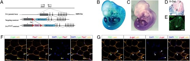
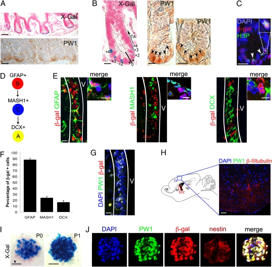
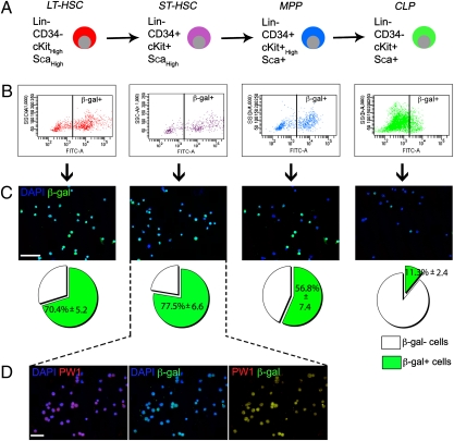
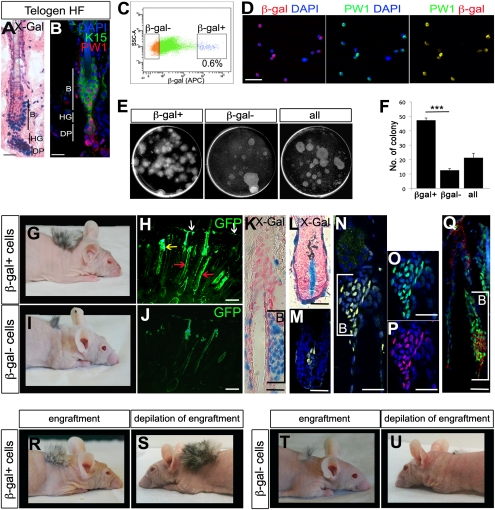
A variety of markers are invaluable for identifying and purifying stem/progenitor cells. Here we report the generation of a murine reporter line driven by Pw1 that reveals cycling and quiescent progenitor/stem cells in all adult tissues thus far examined, including the intestine, blood, testis, central nervous system, bone, skeletal muscle, and skin. Neurospheres generated from the adult PW1-reporter mouse show near 100% reporter-gene expression following a single passage. Furthermore, epidermal stem cells can be purified solely on the basis of reporter-gene expression. These cells are clonogenic, repopulate the epidermal stem-cell niches, and give rise to new hair follicles. Finally, we demonstrate that only PW1 reporter-expressing epidermal cells give rise to follicles that are capable of self-renewal following injury. Our data demonstrate that PW1 serves as an invaluable marker for competent self-renewing stem cells in a wide array of adult tissues, and the PW1-reporter mouse serves as a tool for rapid stem cell isolation and characterization.


Conflict of interest statement

Conflict of interest statement: A patent has been filed for the Tg(Pw1IRESnLacZ) mouse model. This mouse will be freely made available to any and all academic laboratories for noncommercial use.

Figures

Fig. 1.
Fig. 1.
Generation and characterization of Tg(Pw1IRESnLacZ) reporter mice. (A) The 508P6 BAC (180 kb) contains all nine exons (gray boxes) of Pw1. Zim1 is located <40 kb from Pw1 and transcribed in the opposite direction (arrows). An IRESnLacZ-pA (blue) and a floxed (red triangles) kanamycin (Kana, pink) cassette were introduced into the 5′ portion of Pw1 exon 9. The kanamycin cassette was excised before BAC injection. (B and C) Whole-mount X-galactosidase (X-Gal) staining of E11 Tg(Pw1IRESnLacZ) (B) and whole-mount in situ hybridization of E11 wild-type embryos (C). The same expression pattern is seen in the hindbrain (hb), brachial arches (ba), heart (h), apical epidermal region of the fore- (fl), and hindlimb (hl) buds, and somites (so). (D and E) sagittal sections of Tg(Pw1IRESnLacZ) (D) and wild-type (E) embryos at E11. Reporter activity (D) and endogenous PW1 protein (E) are detected in the floor and roof plate of the neural tube (arrows) and the hypaxial domain of the somites (arrowhead). (F and G) Cross sections of Tibialis Anterior muscle from 7 wk-old reporter mice. Reporter activity is detected in satellite cells (yellow arrowheads), identified by Pax7 expression, and PICs (white arrowheads) (F). PW1 and reporter activity colocalize (G, white arrowheads). [Scale bars, 100 μm (D, E), 50 μm (F, G).]
Fig. 2.
Fig. 2.
Reporter activity and PW1 expression identify stem/progenitor cells in the adult small intestine and the CNS. (AC) Representative cross sections of small intestine from 7-wk-old reporter mouse (A Upper, B Left, and C) and wild-type mice (A Lower, B Center and Right) at low (A) and high (B and C) magnification. Histochemical (X-Gal, A and B) and immunocytochemical (PW1, A and B) labeling show that reporter activity and endogenous PW1 protein expression are restricted to the basal crypt. We note that the level of PW1 expression in the crypt cells is weak and diffuse. The cells in the crypt are numbered (0 to +4) and the positions of the transit-amplifying cells are indicated (TA, arrow) (B). (C) Immunofluorescent staining for β-gal and phospho-histone3 (H3P) protein reveals reporter activity in cycling cells (arrowheads). (Inset) Double-positive cell at high magnification. (D) Summary schema of the adult stem cell lineage in the brain: the B cells (GFAP+) give rise to the transit-amplifying type C cells (MASH1+) and then to the neuroblasts that express doublecortin (DCX, type A). (EH) Identification of the cells expressing the reporter gene and PW1 in the brain. (E) Colocalization of β-gal (red) and GFAP, MASH1, and DCX (green), in sagittal sections of the subventricular zone (SVZ, white lines) from a 3-mo-old reporter mouse. Cells at higher magnification are shown on the right. (F) Schematic representation of the percentages of β-gal+ cells in each neural stem/progenitor population. Approximately 90% of the GFAP+ neuronal stem cells express β-gal. The percentage of β-gal+ cells sharply decreases in the more committed (MASH+ and DCX+) progenitor populations. Values represent mean % ± SEM. (G) Colocalization of reporter (β-gal) and PW1 in the SVZ (white lines) V: ventricle. (H) (Right) Schematic representation of the brain showing the area of cortex (Cx, blue rectangle) corresponding to the photomicrograph shown in the left. (Left) Sagittal cryosection of 3-mo-old reporter mouse brain stained for PW1 and β-III-tubulin. PW1 is not detected in differentiated neurons identified by β-III-tubulin expression. Hp, hippocampus; OB, olfactory bulb; St, striatum; SVZ, red rectangle, subventricular zone. (I) X-Gal coloration staining of neurospheres, generated from the SVZ of 2-mo-old reporter mice before (P0) and after (P1) one passage. The number of β-gal+ cells by neurosphere increases after one passage (P1). (J) Colocalization of PW1 (green) and β-gal (red) in nestin-positive neurospheres after one passage. [Scale bars, 20 μm (I), 30 μm (E, G and H), 50 μm (B and C), 150 μm (A).]
Fig. 3.
Fig. 3.
Reporter activity is highest in the primitive hematopoietic stem cell populations from 7-wk-old reporter mice. (A) Four populations were obtained based on cell surface gene expression profiles: LT-HSC and ST-HSC, MPP, and CLP. (B and C) Each population was sorted based on β-gal expression (B). Reporter expression was confirmed by immunofluorescence (C) and the percentage of β-gal+ cells was counted for each populations (C, Lower). Reporter activity is higher in the stem cell populations and decreases in the progenitors. Values represent mean % ± SEM. (D) Immunostaining for PW1 and β-gal in the purified ST-HSC population shows a complete colocalization. [Scale bars, 50 μm (C and D).]
Fig. 4.
Fig. 4.
PW1+ cell population corresponds to competent and self-renewing hair-follicle stem cells. (A) Histochemical staining of longitudinal sections of 7-wk-old reporter mice hair follicle (HF) in telogen. Reporter activity is detected in the stem cells compartment of the skin. B, bulge; DP, dermal papilla; HG, hair germ. (B) Representative longitudinal section of 7-wk-old wild-type mouse hair follicle (HF) in telogen stained for PW1 (red) and K15 (green) to identify the bulge (B). As in the case of the reporter, PW1 expression is restricted to the bulge (B), hair germ (HG), and dermal papilla (DP). (C) Single-cell preparations from adult (7-wk-old) H2B-EGFP × Tg(Pw1IRESnLacZ) mouse epidermis were FACS-sorted on the basis of β-gal expression. Approximately 0.6% of sorted cells are β-gal+. (D) β-Gal and PW1 in freshly sorted β-gal+ cells. (E) Sorted (β-gal+, β-gal) and unsorted (all) cells were plated for clonogenicity experiment and colonies were stained with Rhodamine after 16 d in culture. (F) Schematic representation of the number of colonies formed by β-gal+, β-gal, and unsorted (all) cells as shown in E per dish. β-Gal+ cells form more colonies compared with β-gal and unsorted cells. Values represent β-gal number of colony ± SEM t test (***P < 0.001). (G and I) Hair growth resulting from β-gal β-gal+ versus β-gal cell grafts in 2-mo-old nude mice. (H and J) Micrographs of graft tissue sections showing GFP expression in the epidermis (white arrow), sebaceous gland (yellow arrow), and hair follicle (red arrows). (K–Q) Photomicrographs showing the bulge (B, brackets; K and N–Q) and dermal papilla (L and M). Longitudinal sections are stained either histochemically (X-Gal) (K and L) or by immunofluorescence for β-gal (M, N, and P), GFP (M–O and Q, green) and K15 (Q, red). M and N are merged images. (R–U) Photographs show the hair growth of β-gal+ (R and S) versus β-gal (T and U) cell grafts before (R and T) and after injury (depilation, S and U). [Scale bars, 10 μm (A and B), 30 μm (D), 5 μm (H and J), 50 μm (K–Q).]

Similar articles

Cited by

References

    1. Fuchs E. The tortoise and the hair: Slow-cycling cells in the stem cell race. Cell. 2009;137:811–819. - PMC - PubMed
    1. Magavi SS, Leavitt BR, Macklis JD. Induction of neurogenesis in the neocortex of adult mice. Nature. 2000;405:951–955. - PubMed
    1. Barker N, et al. Identification of stem cells in small intestine and colon by marker gene Lgr5. Nature. 2007;449:1003–1007. - PubMed
    1. Jaks V, et al. Lgr5 marks cycling, yet long-lived, hair follicle stem cells. Nat Genet. 2008;40:1291–1299. - PubMed
    1. Relaix F, et al. Pw1, a novel zinc finger gene implicated in the myogenic and neuronal lineages. Dev Biol. 1996;177:383–396. - PubMed

Publication types

MeSH terms

Substances

LinkOut - more resources