. 2011 Aug;29(8):1283-93.
doi: 10.1002/stem.680.

Novel stem/progenitor cell population from murine tracheal submucosal gland ducts with multipotent regenerative potential

Affiliations

Novel stem/progenitor cell population from murine tracheal submucosal gland ducts with multipotent regenerative potential

Ahmed E Hegab et al. Stem Cells. 2011 Aug.

Abstract

The airway epithelium is in direct contact with the environment and therefore constantly at risk for injury. Basal cells (BCs) have been found to repair the surface epithelium (SE), but the contribution of other stem cell populations to airway epithelial repair has not been identified. We demonstrated that airway submucosal gland (SMG) duct cells, in addition to BCs, survived severe hypoxic-ischemic injury. We developed a method to isolate duct cells from the airway. In vitro and in vivo models were used to compare the self-renewal and differentiation potential of duct cells and BCs. We found that only duct cells were capable of regenerating SMG tubules and ducts, as well as the SE overlying the SMGs. SMG duct cells are therefore a multipotent stem cell for airway epithelial repair This is of importance to the field of lung regeneration as determining the repairing cell populations could lead to the identification of novel therapeutic targets and cell-based therapies for patients with airway diseases.


Conflict of interest statement

Disclosure of Potential Conflicts of Interest

There are no conflicts of interest.

Figures

Figure 1
Figure 1
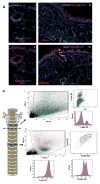
Location of cell types of the cartilaginous airways. (A): Schematic of the airway epithelial cell types of the cartilaginous airways. (B): A few basal cells (BCs) and submucosal gland (SMG) duct cells are the only cells that survive the hypoxic-ischemic injury. Syngeneic heterotopic murine tracheal transplantation was performed, and transplanted tracheas were collected and examined on day 3 when there is maximal cell death. (n = 4–6 tracheas per time point). A longitudinal section of a naïve trachea immunostained with K5, demonstrates the pseudostratified surface epithelium (SE) and the SMG ducts (yellow arrows) of the upper trachea (i). Only SMG duct (white arrows) and a few scattered BCs on the SE survived the hypoxic-ischemic injury. Wide areas of the basement membrane (BM) were denuded and the few cells that remained attached to the BM were K5 positive. SMG ducts were lined with K5+ cells while SMG tubules and myoepithelial cells were necrotic (ii, iii). Annexin V expression was seen in the sloughed cells in the lumen, SMG tubules and ducts (iii). Abbreviation: DAPI: 4′,6-diamidino-2-phenylindole.
Figure 2
Figure 2
Characterization and isolation of submucosal gland (SMG) duct cells. (A): Immunofluorescent staining of SMG duct cells with primary antibodies for specific surface markers. ITGA6 and NGFR are expressed in basal cells (BCs) and are also expressed in the SMG duct (i, ii). EpCAM is present on all epithelial cells of the surface epithelium (SE), SMG tubules, and SMG duct, but TROP-2 is expressed in the cells of the SE and SMG duct, but not in the SMG tubules (iii, iv). K14 is expressed in SMG duct cells, and myoepithelial cells of the tubules (iv). (B): Fluorescence-activated cell sorting of SMG duct and BC populations. (i) The scatter plot shows the distribution of the tracheal cells remaining after stripping of the SE, that is, SMG ducts, tubules and surrounding stroma. TROP-2+ duct cells (blue) and TROP-2- nonduct cells (green) were sorted. Duct cells represented 20% of the total cells. (ii) The scatter plot shows the distribution of the stripped tracheal surface epithelial cells. TROP-2+ITGA6+ BCs (green) and TROP-2+ITGA6- non-BCs (red) were sorted. BCs represented approximately 35% of the total cells. Abbreviations: APC: Allophycocyanin; DAPI: 4′,6-diamidino-2-phenylindole; EpCAM: epithelial cell adhesion molecule; FSC-A: forward scatter; PE: Phycoerythrin; SSC-A : side scatter; TROP-2 : tumor-associated calcium signal transducer 2.
Figure 3
Figure 3
In vitro self-renewal and differentiation ability of submucosal gland (SMG) duct cells compared with basal cells (BCs). (A): The sphere-forming assay: Sphere morphology, clonality, and ability to synthesize mucin. Sorted TROP-2+ SMG duct cells were capable of generating spheres in vitro. Morphologically, two different kinds of spheres were generated; luminal type spheres (i-left) and dense spheres with little or no lumen (i-right). The dense spheres were generally smaller in diameter than the luminal spheres and ranged in size from 20 to 100 μm. The luminal spheres raged from 50 to 300 μm. H&E of a cross-section of the spheres demonstrated the lack of a central lumen in the dense spheres as compared with the luminal spheres (ii). Mixed 1:1 ratio of GFP:wild-type-sorted duct cells demonstrated only green or wild-type spheres, indicating that spheres arose clonally from a single stem/progenitor cell (iii). Sorted duct cells produced mucus in the sphere cultures as seen by Alcian Blue/periodic acid-Schiff (AB/PAS) staining (iv). Sorted TROP-2+integrin-α-6+ BCs from the surface epithelium also generated spheres in vitro. Morphologically, there were just luminal spheres, which varied in size from 50 to 300 μm (v, vi). Mixed 1:1 ratio of GFP:wild-type-sorted BCs demonstrated only green or wild-type spheres, indicating that spheres arose clonally from a single stem/progenitor cell (vii). Sorted BCs produced mucus in the sphere cultures as seen by AB/PAS staining (viii). (B): The sphere-forming assay: Differentiation ability. Dense spheres from duct cells expressed K5 and K14 in all the cells of the sphere (i), with K5 being more strongly expressed on the outside (basal side) of the sphere, with K14 being strongly expressed on the inside (luminal side) of the sphere. K15 was expressed in all cells of the sphere (ii). The dense spheres expressed K8 (48.25% ± 26%) in a luminal pattern (iii). p63 was found in two patterns, either on the basal surface or throughout the dense sphere (iv, v). Spheres expressed the serous cell marker (13.3% ± 5.1%), pIgR (vi). TROP-2 was found on all cells of the sphere (vii). The basal spheres expressed K5 and K14 in almost all the cells of the sphere (viii). p63 was found in cells located on the periphery (xi) and 100% of the spheres expressed K8 in a central pattern (x). K15 was expressed equally in all cells of the sphere (ix). Spheres expressed the secretory marker (56.2% ± 25.2%), pIgR, and 60% ± 11.52% expressed the ciliated marker, acetylated β-tubulin (xii). TROP-2 was found on all cells of the spheres (xiii). Abbreviations: DAPI: 4′,6-diamidino-2-phenylindole; pIgR: polymeric immunoglobulin receptor; TROP-2: tumor-associated calcium signal transducer 2.
Figure 4
Figure 4
Gene expression profiling of submucosal gland (SMG) duct cells compared with basal cells (BCs). (A): The heatmap shows the 100 most differentially expressed genes between sorted duct and BCs and demonstrates that their transcriptional profile is substantially different between the BCs and the duct cells, while their patterns are quite similar within replicates. (B): The ingenuity pathways analysis database was used to analyze biological functions and pathways of the 74 most highly differentially expressed genes. Pathway analysis revealed enrichment in genes in duct cells compared with BCs that were related to epithelial development, such as skin and hair development and dermatological conditions and diseases. (C): Validation of the microarray gene expression of candidate genes was performed with quantitative real-time PCR. K14, K5, Igfbp4, and Asprv1 were more highly expressed in duct as compared with BCs. Data were analyzed using a two-tailed unequal variance Student’s t test. Asterisk indicates a p-value <.005. Dsgl1 trended to significance with a p-value of 0.06. (D): Immunofluorescent staining of IGFBP4 and ASPRV1 in tracheal samples confirmed the location of these proteins was mostly in the SMG duct. ASPRV1 is also located in a few apical cells of the surface epithelium (SE) adjacent to the opening of SMG ducts on the SE. Abbreviations: ALDH1A3: aldehyde dehydrogenase 1A3; ASPRV1: aspartic peptidase, retroviral-like 1; CRYAB: crystallin, alpha B; DAPI: 4′,6-diamidino-2-phenylindole ; DEFB -beta defensin; DIO1: deiodinase, iodothyronine, type I; DSG1: desmoglein 1; ERK1/2: extracellular signal-regulated kinase 1/2; EXP1: exportin 1; FABP5: fatty acid binding protein 5; FCGR1C: Fc fragment of IgG, high affinity Ic, receptor; IGFBP4: insulin-like growth factor binding protein 4; IL1A: interleukin 1, alpha; JNK: c-Jun N-terminal kinase; KRT: keratin; LGALS7: lectin, galactose binding, soluble 7; NFKB: nuclear factor kappa-B; PDGF B: platelet derived growth factor, B polypeptide; SERPINB4: serpin peptidase inhibitor, clade B (ovalbumin), member 4; SLC6A2: solute carrier family 6 (neurotransmitter transporter, noradrenalin), member 2; TG: thyroglobulin.
Figure 5
Figure 5
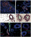
Sorted RFP+TROP-2+ duct cells reconstitute submucosal gland (SMG) tubule-like and SMG duct-like structures in an in vivo regeneration model. To examine the ability of SMG duct cells to reconstitute SMG-like and SMG duct-like structures in vivo, we placed fluorescence-activated cell sorting-sorted RFP+TROP-2+ duct cells into the fat pad under the skin of the mouse back (n = 20, 5,000 cells per mice). We found that in 50% of the grafts, SMG duct cells could organize themselves into SMG tubule-like structures (i–v) and were RFP+, that is, of donor origin (i). These SMG-like structures were K5+ and K14+ (ii, v) and expressed the serous cell marker, lysozyme (vi). In 20% of grafts SMG duct-like structures were seen in addition to the typical tubule-like structures (arrows) (ii). Cells around the SMG tubule-like structures expressed K5 and αSMA, which are markers of myoepithelial cells (v, vi). Abbreviation: αSMA: α-smooth muscle actin.
Figure 6
Figure 6
Lineage tracing of K14-YFP-expressing cells in the in vivo tracheal transplant airway regeneration model after hypoxic-ischemic injury. In naïve mouse tracheas, cells that express K14 are almost entirely located in the submucosal gland (SMG) ducts (white arrow) and thin myoepithelial cells, and only 10% of surface epithelium (SE) basal cells express K14 (green arrow) (i). To trace the contribution of K14+ SMG duct cells to the repair of the airway epithelium after hypoxic-ischemic injury, we used a transgenic mouse model to selectively induce YFP expression in K14-expressing cells. K14CrePR mice [25] were bred with ROSA26-floxed STOP-YFP mice. RU486 or vehicle control was administered intratracheally on day 0. On day 6, tracheas were harvested from these transgenic mice and tracheas were immersed in RU486 or vehicle control, before being transplanted heterotopically into wild-type recipient mice. These tracheal transplants were harvested 7 days later and immunostaining was performed for YFP and K5 and K14 expression. YFP expression was found to colocalize with regions of K14 expression of the SMG, SMG duct and the SE adjacent to and overlying the SMG ducts (ii, iii). In the lower two thirds of the trachea, which does not have SMGs, a few randomly scattered foci of YFP+K14+ cells were seen in the SE (iv, arrow). Abbreviation: GFP: green fluorescent protein.

References

    1. Bartlett JA, Fischer AJ, McCray PB., Jr Innate immune functions of the airway epithelium. Contrib Microbiol. 2008;15:147–163. - PubMed
    1. Schoch KG, Lori A, Burns KA, et al. A subset of mouse tracheal epithelial basal cells generates large colonies in vitro. Am J Physiol Lung Cell Mol Physiol. 2004;286:L631–L642. - PubMed
    1. Hong KU, Reynolds SD, Watkins S, et al. In vivo differentiation potential of tracheal basal cells: Evidence for multipotent and unipotent subpopulations. Am J Physiol Lung Cell Mol Physiol. 2004;286:L643–L649. - PubMed
    1. Hong KU, Reynolds SD, Watkins S, et al. Basal cells are a multipotent progenitor capable of renewing the bronchial epithelium. Am J Pathol. 2004;164:577–588. - PMC - PubMed
    1. Rock JR, Onaitis MW, Rawlins EL, et al. Basal cells as stem cells of the mouse trachea and human airway epithelium. Proc Natl Acad Sci USA. 2009;106:12771–12775. - PMC - PubMed

Publication types

MeSH terms