

Evolution of the blood-brain barrier in newly forming multiple sclerosis lesions
- PMID: 21710622
- PMCID: PMC3143223
- DOI: 10.1002/ana.22472
Evolution of the blood-brain barrier in newly forming multiple sclerosis lesions
Abstract
Objective: Multiple sclerosis (MS) lesions develop around small, inflamed veins. New lesions enhance with gadolinium on magnetic resonance imaging (MRI), reflecting disruption of the blood-brain barrier (BBB). Single time point results from pathology and standard MRI cannot capture the spatiotemporal expansion of lesions. We investigated the development and expansion of new MS lesions, focusing on the dynamics of BBB permeability.
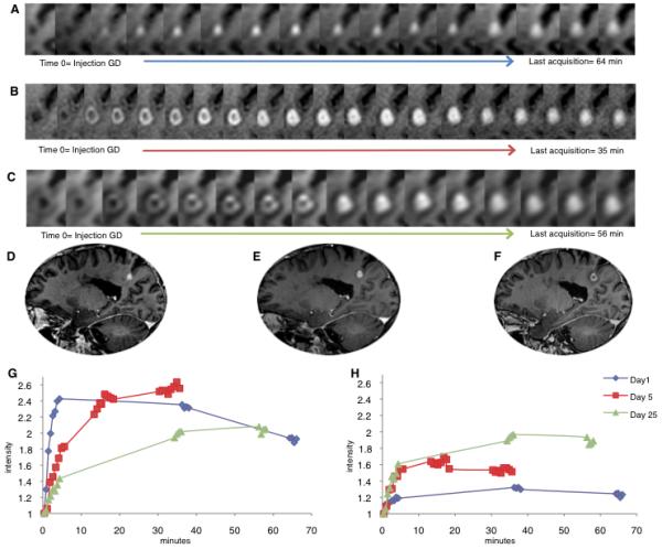
Methods: We performed dynamic contrast-enhanced (DCE) MRI in relapsing-remitting MS. We obtained data over 65 minutes, during and after gadolinium injection. We labeled spatiotemporal enhancement dynamics as centrifugal when initially central enhancement expanded outward and centripetal when initially peripheral enhancement gradually filled the center.
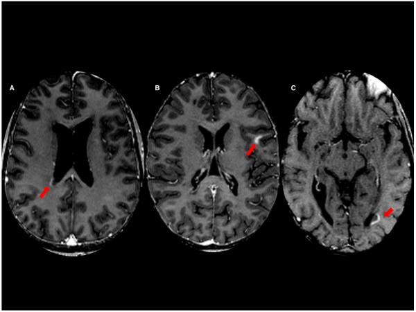
Results: We detected 34 enhancing lesions in 200 DCE-MRI scans. In 65%, enhancement first appeared as a closed ring; in 18%, as a nodule; and in 18%, as an open ring. Lesions with initially nodular enhancement were smaller than those initially enhancing as rings (p < 0.0001). All initially nodular lesions enhanced centrifugally, whereas initially ringlike lesions enhanced centripetally, becoming nodular if small (82%) or nearly nodular if larger (18%). Open-ring lesions were periventricular or juxtacortical and enhanced centripetally. Centrifugally enhancing lesions evolved into centripetally enhancing lesions over several days.
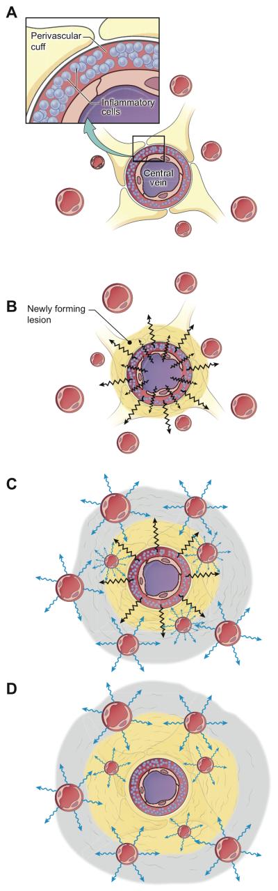
Interpretation: The rapid change of enhancement dynamics from centrifugal to centripetal reflects the outward growth of MS lesions around their central vein and suggests that factors mediating lesion growth and tissue repair derive from different locations at different times. We propose a model of new lesion growth that unites our imaging observations with existing pathology data.
Copyright © 2011 American Neurological Association.
Figures
Comment in
-
A dynamic view of the blood-brain barrier in active multiple sclerosis lesions.Ann Neurol. 2011 Jul;70(1):1-2. doi: 10.1002/ana.22494. Epub 2011 Jun 27. Ann Neurol. 2011. PMID: 21710628 No abstract available.
-
Heterogeneity in longitudinal evolution of ring-enhancing multiple sclerosis lesions.Ann Neurol. 2011 Oct;70(4):668-9; author reply 669-70. doi: 10.1002/ana.22622. Ann Neurol. 2011. PMID: 22028230 Free PMC article. No abstract available.
References
-
- Charcot JM. Histologie de la sclérose en plaques. Gaz des Hôp (Paris) 1868;41:554–66.
-
- Adams CW. The onset and progression of the lesion in multiple sclerosis. J Neurol Sci. 1975 Jun;25(2):165–82. - PubMed
-
- Barnett MH, Prineas JW. Relapsing and remitting multiple sclerosis: pathology of the newly forming lesion. Ann Neurol. 2004 Apr;55(4):458–68. - PubMed
-
- Henderson AP, Barnett MH, Parratt JD, Prineas JW. Multiple sclerosis: distribution of inflammatory cells in newly forming lesions. Ann Neurol. 2009 Dec;66(6):739–53. - PubMed
Publication types
MeSH terms
Grants and funding
LinkOut - more resources
Full Text Sources
Other Literature Sources
Medical
