

Orthotopic xenografts of RCC retain histological, immunophenotypic and genetic features of tumours in patients
- PMID: 21710693
- PMCID: PMC3793840
- DOI: 10.1002/path.2929
Orthotopic xenografts of RCC retain histological, immunophenotypic and genetic features of tumours in patients
Abstract
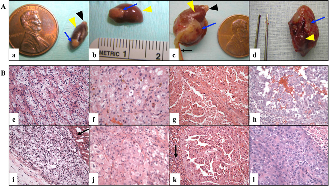
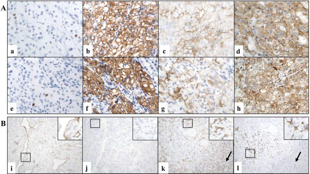
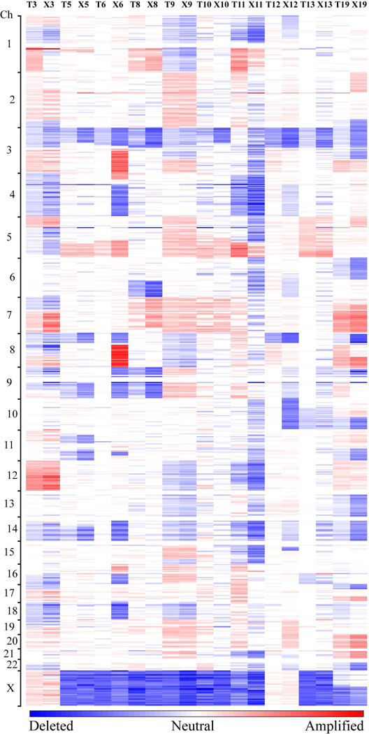
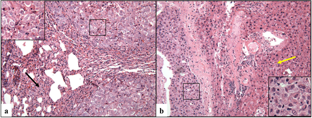
Renal cell carcinoma (RCC) is an aggressive malignancy with limited responsiveness to existing treatments. In vivo models of human cancer, including RCC, are critical for developing more effective therapies. Unfortunately, current RCC models do not accurately represent relevant properties of the human disease. The goal of this study was to develop clinically relevant animal models of RCC for preclinical investigations. We transplanted intact human tumour tissue fragments orthotopically in immunodeficient mice. The xenografts were validated by comparing the morphological, phenotypic and genetic characteristics of the kidney tumour tissues before and after implantation. Twenty kidney tumours were transplanted into mice. Successful tumour growth was detected in 19 cases (95%). The histopathological and immunophenotypic features of the xenografts and those of the original tumours largely overlapped in all cases. Evaluation of genetic alterations in a subset of 10 cases demonstrated that the grafts largely retained the genetic features of the pre-implantation RCC tissues. Indeed, primary tumours and corresponding grafts displayed identical VHL mutations. Moreover, an identical pattern of DNA copy amplification or loss was observed in 6/10 cases (60%). In summary, orthotopic engrafting of RCC tissue fragments can be successfully used to generate animal models that closely resemble RCC in patients. These models will be invaluable for in vivo preclinical drug testing and for deeper understanding of kidney carcinogenesis. The raw data of the SNP array analysis has been submitted to the GEO database (Accession No. GSE29062).
Copyright © 2011 Pathological Society of Great Britain and Ireland. Published by John Wiley & Sons, Ltd.
Conflict of interest statement
The authors declare no conflict of interest.
Figures
References
-
- Cohen HT, McGovern FJ. Renal-cell carcinoma. N Engl J Med. 2005 Dec 8;353(23):2477–2490. - PubMed
-
- Jemal A, Siegel R, Ward E, Hao Y, Xu J, Thun MJ. Cancer statistics, 2009. CA Cancer J Clin. 2009 Jul-Aug;59(4):225–249. - PubMed
-
- McLaughlin JK, Lipworth L, Tarone RE. Epidemiologic aspects of renal cell carcinoma. Semin Oncol. 2006 Oct;33(5):527–533. - PubMed
-
- Rini BI, Campbell SC, Escudier B. Renal cell carcinoma. Lancet. 2009 Mar 28;373(9669):1119–1132. - PubMed
-
- Hollingsworth JM, Miller DC, Daignault S, Hollenbeck BK. Rising incidence of small renal masses: a need to reassess treatment effect. J Natl Cancer Inst. 2006 Sep 20;98(18):1331–1334. - PubMed
Publication types
MeSH terms
Associated data
- Actions
Grants and funding
LinkOut - more resources
Full Text Sources
Other Literature Sources
Medical
Molecular Biology Databases
