. 2011 Aug 4;115(30):9495-506.
doi: 10.1021/jp204787b. Epub 2011 Jul 13.

Carotenoid radical formation: dependence on conjugation length

Affiliations

Carotenoid radical formation: dependence on conjugation length

A Ligia Focsan et al. J Phys Chem B. .

Abstract

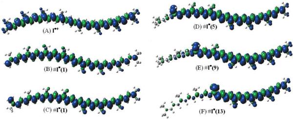
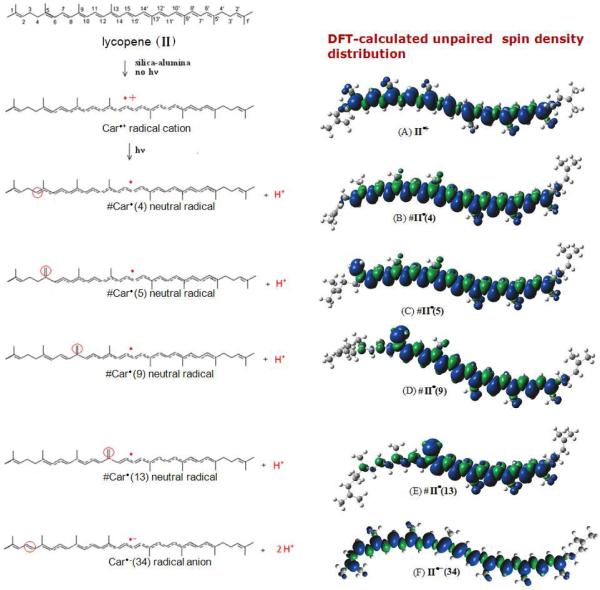
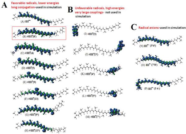
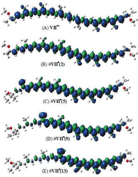
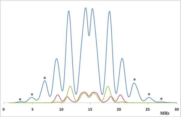
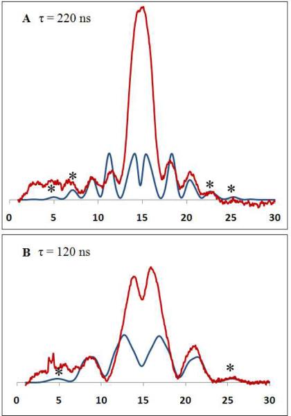
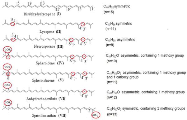
The relative energy of carotenoid neutral radicals formed by proton loss from the radical cations of linear carotenoids has been examined as a function of conjugation length from n = 15 to 9. For a maximum conjugation length of n = 15 (bisdehydrolycopene, a symmetrical compound), proton loss can occur from any of the 10 methyl groups, with proton loss from the methyl group at position C1 or C1' being the most favorable. In contrast, the most energetically favorable proton loss from the radical cations of lycopene, neurosporene, spheroidene, spheroidenone, spirilloxanthin, and anhydrorhodovibrin occurs from methylene groups that extend from the conjugated system. For example, decreasing the conjugation length to n = 11 (lycopene) by saturation of the double bonds C3-C4 and at C3'-C4' of bisdehydrolycopene favors proton loss at C4 or C4' methylene groups. Saturation at C7'-C8' in the case of neurosporene, spheroidene, and spheroidenone (n = 9, 10, 11) favors the formation of a neutral radical at the C8' methylene group. Saturation of C1-C2 by addition of a methoxy group to a bisdehydrolycopene-like structure with conjugation of n = 12 or 13 (anhydrorhodovibrin, spirilloxanthin) favors proton loss at the C2 methylene group. As a consequence of deprotonation of the radical cation, the unpaired electron spin distribution changes so that larger β-methyl proton couplings occur for the neutral radicals (13-16 MHz) than for the radical cation (7-10 MHz), providing a means to identify possible carotenoid radicals in biological systems by Mims ENDOR.


Figures

Figure 1
Figure 1
Unpaired spin density distribution for bisdehydrolycopene radicals (A) I•+, (B) #I(1) cis, (C) #I(1) trans, (D) #I(5), (E) #I(9) and (F) #I(13) from DFT calculations.
Figure 2
Figure 2
Possible intermediates for lycopene (II); Unpaired spin density distribution for lycopene radicals (A) II•+, (B) #II(4), (C) #II(5), (D) #II(9), (E) #II(13) and (F) II•− (34) from DFT calculations.
Figure 3
Figure 3
Unpaired spin density distribution for neurosporene (A) III•+, (B) #III(8'), (C) #III(4), (D) #III(5), (E) #III(9), (F) #III(13), (G) #III(9'), (H) #III(13'), (I) #III(3), (J) #III(3') (K) #III(4'), (L) #III(5'), (M) #III(7'), (N) III•−(3'4') (O) III•−(7'8') and (P)(III•−(34) from DFT calculations.
Figure 4
Figure 4
Unpaired spin density distribution for spheroidene radicals (A) IV•+, (B) IV(2), (C) #IV(5), (D) #IV(9), (E) #IV(13), (F) #IV(3'), (G) #IV(4'), (H) #IV(5'), (I) #IV(7'), (J) #IV(8'), (K) #IV(9') and (L) #IV(13') from DFT calculations.
Figure 5
Figure 5
Unpaired spin density distribution for spheroidenone radicals (A)V•+, (B) #V(5), (C) #V(9), (D) #V(13), (E) #V(3'), (F) #V(4'), (G) #V(5'), (H) #V(7'), (I) #V(8'), (J) #V(9') and (K) #V(13') from DFT calculations.
Figure 6
Figure 6
Unpaired spin density distribution for anhydrorhodovibrin radicals (A)VI•+, (B) #VI(2), (C) #VI(5), (D) #VI(9), (E) #VII(13), (F) #VI(3'), (G) #VI(4'), (H) #VI(5'), (I) #VI(9') and (J) #VI(13') from DFT calculations.
Figure 7
Figure 7
Unpaired spin density distribution for spirilloxanthin radicals (A)VII•+, (B) #VII(2),(C) #VII(5), (D) #VII(9) and (E) #VII(13) from DFT calculations.
Figure 8
Figure 8
The relative energies (ΔE(n) in kcal/mol, Table 1) for loss of a proton from the radical cations versus relative change in delocalization N-Nmax for I–VII. Smooth line has been drawn as a guide for the eye. Note: The localized neutral radicals having high relative energy and short conjugated chain (~4 C atoms) were not included.
Figure 9
Figure 9
A) Individual contribution of spirilloxanthin radicals (VII) to the Mims ENDOR spectrum; VII•+ is indicated by the grey shaded area, #VII(2) by red, #VII(5) by green, #VII(9) by purple and #VII(13) by blue lines. B) Simulated Mims ENDOR spectrum of the sum of spirilloxanthin radicals VII•+, #VII(2), #VII(5), #VII(9) and #VII(13) in 1:1:1:1:1 ratio. Note: the outer peaks indicated by * are due to the neutral radicals.
Figure 10
Figure 10
Simulated Mims ENDOR spectrum of neurosporene (III) radicals (in blue) τ = 220 ns, B = 3475 G, mw = 9.76 GHz, νn = 14.79 MHz using the hyperfine coupling tensors given in Supporting Information. The blue plot is the simulation of III•+, III•−(34), III•−(3'4'), III•−(7'8'), #III(4), #III(5), #III(9), #III(13), #III(8'), #III(9'), #III(13') radicals in equal amounts. The red plot is the simulation of the radical cation III•+; the green plot is the simulation of the radical anions III•−(34), III•−(3'4') and III•−(7'8'). Note: the outer peaks indicated by * are due to the neutral radicals.
Figure 11
Figure 11
Pulsed Mims ENDOR spectra of lycopene (II) radicals as a function of τ. A) 220 ns and B) 120 ns. The red trace is the experimental spectrum produced in activated silica-alumina after UV irradiation. ENDOR parameters: T = 20 K, B = 3475 G, ν = 9.764 GHz. The blue trace is the simulated spectrum using isotropic and anisotropic DFT-calculated couplings listed in Tables S7–S11 Supporting Information for II•+, #II(4) #II(5) #II(9) and #II•(13) in a 1:1:1:1:1 ratio. Note: ENDOR lines occur at νnA/2, where νn is the proton frequency situated at the center of the ENDOR spectrum (proton frequency νn = 14.793571 MHz). Note: the outer peaks indicated by * are due to the neutral radicals. Below 6 MHz the baseline includes an artifact from the nonlinearity of the ENDOR amplifier and thus interferes with low-frequency ENDOR lines due to neutral radicals.
Scheme 1
Scheme 1
Linear carotenoids

References

    1. Koepke J, Hu X, Muenke C, Schulten K, Michel H. Structure. 1996;4:581–97. - PubMed
    1. Qian P, Saiki K, Mizoguchi T, Hara K, Sashima T, Fujii R, Koyama Y. Photochem Photobiol. 2001;74:444–52. - PubMed
    1. Yeliseev AA, Eraso JM, Kaplan S. J Bacteriol. 1996;178:5877–83. - PMC - PubMed
    1. Polivka T, Pullerits T, Frank HA, Cogdell RJ, Sundstrom V. J Phys Chem B. 2004;108:15398–15407.
    1. Wormit M, Harbach PH, Mewes JM, Amarie S, Wachtveitl J, Dreuw A. Biochim Biophys Acta. 2009;1787:738–46. - PubMed

Publication types