. 2011 Sep;134(1):50-9.
doi: 10.1111/j.1365-2567.2011.03466.x. Epub 2011 Jun 29.

Induction of self-antigen-specific Foxp3+ regulatory T cells in the periphery by lymphodepletion treatment with anti-mouse thymocyte globulin in mice

Affiliations

Induction of self-antigen-specific Foxp3+ regulatory T cells in the periphery by lymphodepletion treatment with anti-mouse thymocyte globulin in mice

Yan Lu et al. Immunology. 2011 Sep.

Abstract

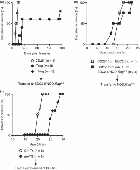
Lymphodepletion therapies are increasingly tested for controlling immune damage. One appealing premise for such a therapy is that it may 'reboot' the immune system and restore immune tolerance. However, the tolerogenic potential of lymphodepletion therapies remains controversial. The debate is exemplified by conflicting evidence from the studies of anti-thymocyte globulin (ATG), a prototype of immunodepleting drugs, in particular on whether it induces CD4(+) CD25(+) Foxp3(+) regulatory T (Treg) cells. To understand the impact of ATG on T cells at a clonal level in vivo, we studied the effect of anti-mouse thymocyte globulin (mATG) in a reductionist model in which the T-lymphocyte repertoire consists of a single clone of pathogenic T effector (Teff) cells specific to a physiological self-antigen. The mATG treatment led to peripheral induction of antigen-specific Treg cells from an otherwise monoclonal Teff repertoire, independent of thymic involvement. The de novo induction of Treg cells occurred consistently in local draining lymph nodes, and persistence of induced Treg cells in blood correlated with long-term protection from autoimmune destruction. This study provides in vivo evidence for clonal conversion from a pathogenic self-antigen-specific Teff cell to a Treg cell in the setting of immunodepletion therapies.


Figures

Figure 1
Figure 1
Lymphodepletion by anti-mouse thymocyte globulin (mATG) inhibited autoimmunity with a gender effect. The mATG was administered to female or male BDC2.5/NOD.Rago/o mice, which harbour a monoclonal T-cell repertoire consisting of a single pathogenic effector T (Teff) -cell clone specific to a self-antigen in pancreatic beta cells. The effect of mATG on the T-cell-mediated autoimmune damage was assessed by development of autoimmune diabetes. (a) The effect of different doses of mATG. (b) The effect of mATG treatment in female mice compared with that in male animals. Graph represents 4–10 mice in each group.
Figure 2
Figure 2
Anti-mouse thymocyte globulin (mATG) depleted peripheral T lymphocytes but not thymocytes. The mATG-treated BDC2.5/NOD.Rago/o mice were analysed at the time of active mATG-mediated depletion (within 2–3 days after the last mATG dose) (middle panels, mATG depletion), or 3 weeks after mATG treatment, when the T cells recovered (bottom panels, post-mATG), in comparison to pre-diabetic control mice treated with rabbit IgG or that received no treatment around 3 weeks of age (top panels). MLN, mesenteric lymph nodes; PLN, pancreatic draining lymph nodes. (a) Plots are derived from a lymphoid cell gate in forward versus side scatter plots. Numbers in the graphs are percentages of the gated populations. (b) Statistical representations (mean ± SD) of five to seven animals in each group, including at least two animals in either gender (no gender difference was observed in the extent of depletion). *P < 0·005 (Student's t-test comparing mATG treated versus control groups).
Figure 3
Figure 3
Local induction of CD4+ Foxp3+ CTLA4+ regulatory T cells in the periphery from a monoclonal Teff-cell repertoire. anti-mouse thymocyte globulin (mATG) -treated BDC2.5/NOD.Rago/o mice were analysed 3 weeks after the depletion treatment (bottom panels), when the T lymphocytes repopulated, in comparison to control mice treated with the same dose of rabbit IgG or with no treatment (top panels) (n = 5 for each group). Control mice were used at about 3 weeks of age (before diabetes occurred). MLN, mesenteric lymph nodes; PLN, pancreatic draining lymph nodes. (a) Numbers in the plots are the percentages of the delineated populations. Gate, CD4+ CD8 cells. (b) Statistical representations (mean ± SD) of five animals in each group, including at least two animals in either gender. *P < 0·0001 (Student's t-test).
Figure 4
Figure 4
Appearance of induced Foxp3+ regulatory T (Treg) cells in peripheral blood correlated with induction of immune tolerance in a model of aggressive autoimmune damage. (a) A representative profile of Foxp3 expression by induced Treg cells (iTreg) in peripheral blood of Rag-deficient BDC2.5 mice versus natural Treg cells (nTreg) from Rag-sufficient BDC2.5 mice. (b) Percentage of Foxp3+ cells among CD4+ T cells in the peripheral blood of the anti-thymocyte globulin (ATG) -treated male (open or closed triangle) or female (open circle) mice, grouped based on length of diabetes-free time. Each symbol represents one animal. The iTreg cells appeared in high percentages in the blood of a fraction of male animals treated with mATG. (c) Kinetics of iTreg cells in peripheral blood of the animals protected for > 160 days. Each line represents one animal.
Figure 5
Figure 5
The induced Treg cells in the peripheral blood of anti-mouse thymocyte globulin (mATG) -treated, long-lived BDC2.5/NOD.Rago/o male mice expressed higher levels of CD103. Blood samples from the mATG-protected animals (right) were analysed by flow cytometry, in comparison to that of BDC2.5/NOD mice. Gated on CD4+ Foxp3+ (top panels, regulatory T cells) or CD4+ Foxp3 (bottom panels, effector T cells). Numbers in the plots are the percentages of the delineated populations.
Figure 6
Figure 6
Essential role of induced Foxp3+ regulatory T (Treg) cells for the long-term protective effect of anti-mouse thymocyte globulin (mATG). (a) Adoptive transfer experiments to test the efficacy of mATG-induced Treg (iTreg) cells from long-lived, male BDC2.5/NOD.Rago/o mice. CD4+ T cells from mATG-treated BDC2.5/NOD.Rago/o mice were fractionated into CD25+ (iTreg) or CD25 subsets by flow cytometry sorting and transferred into new BDC2.5/NOD.Rago/o recipients at the age of 8–13 days (7 × 10⁴ cells for each recipient), to examine their potential suppressive activity on autoimmune damage, in comparison to natural Treg (nTreg) cells from unmanipulated BDC2.5/NOD mice. (b) Adoptive transfer experiments to test the pathogenic effect of CD4+ CD25 cells in mATG-tolerized BDC2.5/NOD.Rago/o mice. CD4+ CD25 cells were sorted from mATG-treated BDC2.5/NOD.Rago/o mice by flow cytometry and transferred into Rago/o recipients (7 × 10⁴ cells for each recipient), to examine their autoimmune pathogenic activity in comparison to the activity of CD4+ CD25 Teff cells from unmanipulated BDC2.5/NOD mice. (c) To examine Foxp3-independent activity, 20 mg/kg mATG or control treatment were administered to male Foxp3-deficient BDC2.5 mice (BDC2.5/NOD.Foxp3sf) using the same schedule of treatment as in Fig. 1 for BDC2.5/NOD.Rago/o mice, beginning at 10 days of age, once every 4 days, for four doses.

References

    1. Sakaguchi S, Yamaguchi T, Nomura T, Ono M. Regulatory T cells and immune tolerance. Cell. 2008;133:775–87. - PubMed
    1. Beiras-Fernandez A, Thein E, Hammer C. Induction of immunosuppression with polyclonal antithymocyte globulins: an overview. Exp Clin Transplant. 2003;1:79–84. - PubMed
    1. Mohty M, Gaugler B. Mechanisms of action of antithymocyte globulin: old dogs with new tricks! Leuk Lymphoma. 2008;49:1664–7. - PubMed
    1. Lopez M, Clarkson MR, Albin M, Sayegh MH, Najafian N. A novel mechanism of action for anti-thymocyte globulin: induction of CD4+ CD25+ Foxp3+ regulatory T cells. J Am Soc Nephrol. 2006;17:2844–53. - PubMed
    1. Feng X, Kajigaya S, Solomou EE, et al. Rabbit ATG but not horse ATG promotes expansion of functional CD4+ CD25high FOXP3+ regulatory T cells in vitro. Blood. 2008;111:3675–83. - PMC - PubMed

Publication types

MeSH terms

LinkOut - more resources