. 2011 Jun 27;1(1):23.
doi: 10.1186/2045-3701-1-23.

TBP-related factors: a paradigm of diversity in transcription initiation

Affiliations

TBP-related factors: a paradigm of diversity in transcription initiation

Waseem Akhtar et al. Cell Biosci. .

Abstract

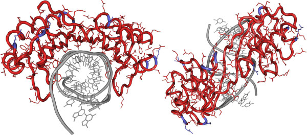
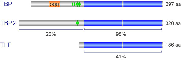
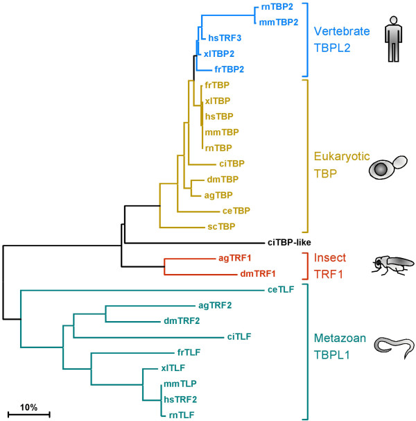
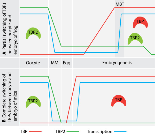
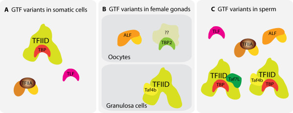
TATA binding protein (TBP) is a key component of the eukaryotic transcription initiation machinery. It functions in several complexes involved in core promoter recognition and assembly of the pre-initiation complex. Through gene duplication eukaryotes have expanded their repertoire of TATA binding proteins, leading to a variable composition of the transcription machinery. In vertebrates this repertoire consists of TBP, TBP-like factor (TLF, also known as TBPL1, TRF2) and TBP2 (also known as TBPL2, TRF3). All three factors are essential, with TLF and TBP2 playing important roles in development and differentiation, in particular gametogenesis and early embryonic development, whereas TBP dominates somatic cell transcription. TBP-related factors may compete for promoters when co-expressed, but also show preferential interactions with subsets of promoters. Initiation factor switching occurs on account of differential expression of these proteins in gametes, embryos and somatic cells. Paralogs of TFIIA and TAF subunits account for additional variation in the transcription initiation complex. This variation in core promoter recognition accommodates the expanded regulatory capacity and specificity required for germ cells and embryonic development in higher eukaryotes.


Figures

Figure 1
Figure 1
Structure of core domain of human TBP bound to the TATA element of the adenovirus major late promoter. The DNA is shown in grey, TBP is shown in red. Amino acids that are different between TBP and TBP2 are shown in blue. Left: Frontal view, showing that all amino acids contacting the DNA are identical between TBP and TBP2 (red). Substituted amino acids are solvent exposed (blue). Right: Top view, showing that TBP aligns with the minor groove in which it is inserted.
Figure 2
Figure 2
Domain structure of the vertebrate TBP family. The core domain (blue) is formed by an imperfect direct repeat (blue) and is involved in binding to the TATA box in the case of TBP and TBP2. TLF lacks an N-terminal domain. The N-terminal domain of TBP2 is not as well conserved as the core domain. The PXT repeats in the N-terminal domain are indicated (green), as well as the poly-glutamine stretch of TBP (orange).
Figure 3
Figure 3
Phylogenetic tree of the TBP-related factors in eukaryotes based on alignment of their core domains. TBP (gold) is the founding member of the family with representatives in archae (not shown), yeast and all metazoans. TRF (TRF1, red) is found in the fruit fly and the mosquito. TLF (TBPL1, TRF2, light green) is found in all multicellular organisms, but not in yeast. TBP2 (TBPL2, TRF3, blue) is restricted to vertebrates. The bar signifies the distance in the tree corresponding to 10% divergence. Legend: hs-Homo sapiens, mm-Mus musculus, rn-Rattus norvegicus, xl-Xenopus laevis, fr-Fugu rubripes, ci-Ciona intestinalis, dm-Drosophila melanogaster, ag-Anopheles gambiae, ce-Caenorhabditis elegans, sc-Saccharomyces cerevisiae. ciTBP: XP_002127120, ciTLF: XP_002128047, ciTBP-like: BW175023, agTBP: XP_309748, agTRF1: XP_317373, agTRF2: XP_308361.
Figure 4
Figure 4
Switching of TATA binding proteins between oocytes and embryos. (A) In Xenopus oocytes TBP2 but not TBP is present at the protein level. Upon meiotic maturation (MM) TBP2 is degraded after global shut down of transcription, but residual levels of TBP2 persist in egg and early embryo. During early cleavages after fertilization, maternal stores of TBP mRNA are translated and by the mid-blastula transition (MBT) both TBP and residual TBP2 contribute to zygotic transcription leading to a partial switching of function between TBP and TBP2. In X. laevis TBP2 RNA is detected well into larval stages, whereas in X. tropicalis TBP2 mRNA levels decline during gastrulation (our unpublished data). (B) Mouse oocytes also express only TBP2. When zygotic transcription commences at the two cell stage of the mouse embryo, TBP2 function is replaced by translationally upregulated TBP leading to a complete switching of TATA binding factors.
Figure 5
Figure 5
Many germ cell-specific variants of general transcription factors (GTFs) have evolved. Only factors are shown for which a role has been documented. While TBP dominates transcription in somatic cells (A) and is also expressed in testis (C), it is completely replaced by TBP2 in female germ cells (B). Male germ cells express high levels of TLF and a vital share of transcription in these cells is dependent on this factor. Similarly TFIIA is replaced completely in oocytes and partially in sperm by ALF. Variants of TFIID contain germ cell-specific TAFs.

References

    1. Juven-Gershon T, Kadonaga JT. Regulation of gene expression via the core promoter and the basal transcriptional machinery. Dev Biol. pp. 225–229. - PMC - PubMed
    1. Thomas MC, Chiang CM. The general transcription machinery and general cofactors. Crit Rev Biochem Mol Biol. 2006;41(3):105–178. doi: 10.1080/10409230600648736. - DOI - PubMed
    1. Muller F, Tora L. The multicoloured world of promoter recognition complexes. EMBO J. 2004;23(1):2–8. doi: 10.1038/sj.emboj.7600027. - DOI - PMC - PubMed
    1. Hernandez N. TBP, a universal eukaryotic transcription factor? Genes Dev. 1993;7(7B):1291–1308. doi: 10.1101/gad.7.7b.1291. - DOI - PubMed
    1. Burley SK. The TATA box binding protein. Curr Opin Struct Biol. 1996;6(1):69–75. doi: 10.1016/S0959-440X(96)80097-2. - DOI - PubMed