Granulocyte colony stimulating factor attenuates inflammation in a mouse model of amyotrophic lateral sclerosis
- PMID: 21711557
- PMCID: PMC3146845
- DOI: 10.1186/1742-2094-8-74
Granulocyte colony stimulating factor attenuates inflammation in a mouse model of amyotrophic lateral sclerosis
Abstract
Background: Granulocyte colony stimulating factor (GCSF) is protective in animal models of various neurodegenerative diseases. We investigated whether pegfilgrastim, GCSF with sustained action, is protective in a mouse model of amyotrophic lateral sclerosis (ALS). ALS is a fatal neurodegenerative disease with manifestations of upper and lower motoneuron death and muscle atrophy accompanied by inflammation in the CNS and periphery.
Methods: Human mutant G93A superoxide dismutase (SOD1) ALS mice were treated with pegfilgrastim starting at the presymptomatic stage and continued until the end stage. After long-term pegfilgrastim treatment, the inflammation status was defined in the spinal cord and peripheral tissues including hematopoietic organs and muscle. The effect of GCSF on spinal cord neuron survival and microglia, bone marrow and spleen monocyte activation was assessed in vitro.
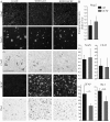
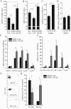
Results: Long-term pegfilgrastim treatment prolonged mutant SOD1 mice survival and attenuated both astro- and microgliosis in the spinal cord. Pegfilgrastim in SOD1 mice modulated the inflammatory cell populations in the bone marrow and spleen and reduced the production of pro-inflammatory cytokine in monocytes and microglia. The mobilization of hematopoietic stem cells into the circulation was restored back to basal level after long-term pegfilgrastim treatment in SOD1 mice while the storage of Ly6C expressing monocytes in the bone marrow and spleen remained elevated. After pegfilgrastim treatment, an increased proportion of these cells in the degenerative muscle was detected at the end stage of ALS.
Conclusions: GCSF attenuated inflammation in the CNS and the periphery in a mouse model of ALS and thereby delayed the progression of the disease. This mechanism of action targeting inflammation provides a new perspective of the usage of GCSF in the treatment of ALS.
Figures


Similar articles
-
Granulocyte Colony-Stimulating Factor Ameliorates Skeletal Muscle Dysfunction in Amyotrophic Lateral Sclerosis Mice and Improves Proliferation of SOD1-G93A Myoblasts in vitro.Neurodegener Dis. 2017;17(1):1-13. doi: 10.1159/000446113. Epub 2016 Aug 20. Neurodegener Dis. 2017. PMID: 27544379
-
Granulocyte-colony stimulating factor improves outcome in a mouse model of amyotrophic lateral sclerosis.Brain. 2008 Dec;131(Pt 12):3335-47. doi: 10.1093/brain/awn243. Epub 2008 Oct 3. Brain. 2008. PMID: 18835867 Free PMC article.
-
Toll-Like Receptor-4 Inhibitor TAK-242 Attenuates Motor Dysfunction and Spinal Cord Pathology in an Amyotrophic Lateral Sclerosis Mouse Model.Int J Mol Sci. 2017 Aug 1;18(8):1666. doi: 10.3390/ijms18081666. Int J Mol Sci. 2017. PMID: 28763002 Free PMC article.
-
Progesterone and Allopregnanolone Neuroprotective Effects in the Wobbler Mouse Model of Amyotrophic Lateral Sclerosis.Cell Mol Neurobiol. 2022 Jan;42(1):23-40. doi: 10.1007/s10571-021-01118-y. Epub 2021 Jun 17. Cell Mol Neurobiol. 2022. PMID: 34138412 Free PMC article. Review.
-
The Role of Skeletal Muscle in Amyotrophic Lateral Sclerosis.Brain Pathol. 2016 Mar;26(2):227-36. doi: 10.1111/bpa.12350. Brain Pathol. 2016. PMID: 26780251 Free PMC article. Review.
Cited by
-
Immunological aspects in amyotrophic lateral sclerosis.Transl Stroke Res. 2012 Sep;3(3):331-40. doi: 10.1007/s12975-012-0177-6. Epub 2012 May 3. Transl Stroke Res. 2012. PMID: 24323808
-
Intranasal Delivery: Effects on the Neuroimmune Axes and Treatment of Neuroinflammation.Pharmaceutics. 2020 Nov 20;12(11):1120. doi: 10.3390/pharmaceutics12111120. Pharmaceutics. 2020. PMID: 33233734 Free PMC article. Review.
-
Cytokine therapy-mediated neuroprotection in a Friedreich's ataxia mouse model.Ann Neurol. 2017 Feb;81(2):212-226. doi: 10.1002/ana.24846. Ann Neurol. 2017. PMID: 28009062 Free PMC article.
-
Early systemic granulocyte-colony stimulating factor treatment attenuates neuropathic pain after peripheral nerve injury.PLoS One. 2012;7(8):e43680. doi: 10.1371/journal.pone.0043680. Epub 2012 Aug 24. PLoS One. 2012. PMID: 22937076 Free PMC article.
-
Are Circulating Cytokines Reliable Biomarkers for Amyotrophic Lateral Sclerosis?Int J Mol Sci. 2019 Jun 5;20(11):2759. doi: 10.3390/ijms20112759. Int J Mol Sci. 2019. PMID: 31195629 Free PMC article. Review.
References
-
- Rosen DR, Siddique T, Patterson D, Figlewicz DA, Sapp P, Hentati A, Donaldson D, Goto J, O'Regan JP, Deng HX, Rahmani Z, Krizus A, McKenna-Yasek D, Cayabyab A, Gaston SM, Berger R, Tanzi RE, Halperin JJ, Herzfeldt B, Van den Bergh R, Hung W, Bird T, Deng G, Mulder DW, Smyth C, Laing NG, Soriano E, Pericak-Vance MA, Haines J, Rouleau GA, Gusella JS, Horvitz HR, Brown RH Jr. Mutations in Cu/Zn superoxide dismutase gene are associated with familial amyotrophic lateral sclerosis. Nature. 1993;362:59–62. doi: 10.1038/362059a0. - DOI - PubMed
-
- Pasinelli P, Brown RH. Molecular biology of amyotrophic lateral sclerosis: insights from genetics. Nat Rev Neurosci. 2006;7:710–723. - PubMed
-
- Jang YC, Lustgarten MS, Liu Y, Muller FL, Bhattacharya A, Liang H, Salmon AB, Brooks SV, Larkin L, Hayworth CR, Richardson A, Van Remmen H. Increased superoxide in vivo accelerates age-associated muscle atrophy through mitochondrial dysfunction and neuromuscular junction degeneration. FASEB J. 2010;24:1376–1390. doi: 10.1096/fj.09-146308. - DOI - PMC - PubMed
Publication types
MeSH terms
Substances
LinkOut - more resources
Full Text Sources
Other Literature Sources
Medical
Miscellaneous

