. 2011 Aug;17(8):1489-501.
doi: 10.1261/rna.2442211. Epub 2011 Jun 28.

A role for noncanonical microRNAs in the mammalian brain revealed by phenotypic differences in Dgcr8 versus Dicer1 knockouts and small RNA sequencing

Affiliations

A role for noncanonical microRNAs in the mammalian brain revealed by phenotypic differences in Dgcr8 versus Dicer1 knockouts and small RNA sequencing

Joshua E Babiarz et al. RNA. 2011 Aug.

Abstract

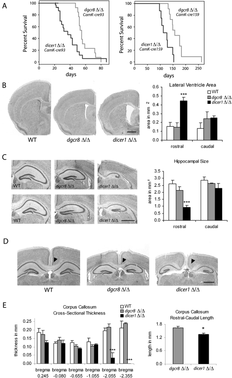
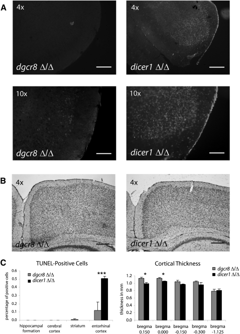
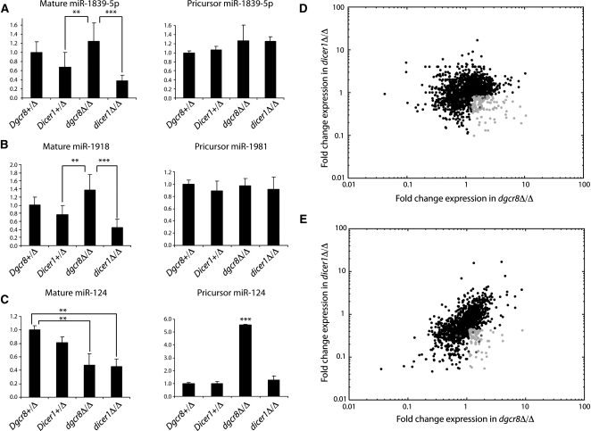
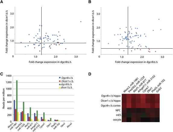
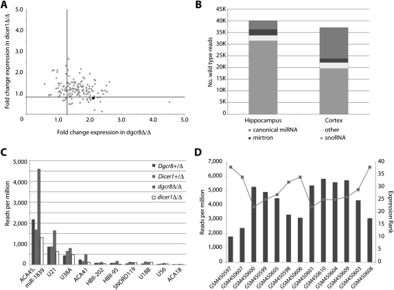
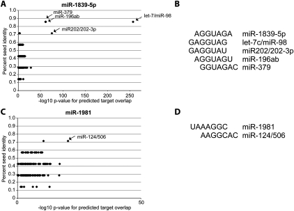
Noncanonical microRNAs (miRNAs) and endogenous small interfering RNAs (endo-siRNAs) are distinct subclasses of small RNAs that bypass the DGCR8/DROSHA Microprocessor but still require DICER1 for their biogenesis. What role, if any, they have in mammals remains unknown. To identify potential functional properties for these subclasses, we compared the phenotypes resulting from conditional deletion of Dgcr8 versus Dicer1 in post-mitotic neurons. The loss of Dicer1 resulted in an earlier lethality, more severe structural abnormalities, and increased apoptosis relative to that from Dgcr8 loss. Deep sequencing of small RNAs from the hippocampus and cortex of the conditional knockouts and control littermates identified multiple noncanonical microRNAs that were expressed at high levels in the brain relative to other tissues, including mirtrons and H/ACA snoRNA-derived small RNAs. In contrast, we found no evidence for endo-siRNAs in the brain. Taken together, our findings provide evidence for a diverse population of highly expressed noncanonical miRNAs that together are likely to play important functional roles in post-mitotic neurons.


Figures

FIGURE 1.
FIGURE 1.
Conditional loss of Dicer1 in post-mitotic neurons resulted in more severe phenotypes than loss of Dgcr8. (A) Survival analysis was performed on CamK-cre93 dgcr8Δ/Δ and dicer1Δ/Δ conditional mice. P-value was determined by the log-rank test. (B) dicer1Δ/Δ animals showed increases in lateral ventricle size at rostral (bregma −0.080, P = 0.0009) but not at caudal (bregma −1.255, P = 0.94) regions. (C) The rostral (bregma −2.055, P = 0.008), but not caudal (bregma −2.48, P = 0.371), hippocampal structure was malformed and reduced in size in the dicer1Δ/Δ brains compared with dgcr8Δ/Δ. (D,E) dicer1Δ/Δ animals exhibited agenesis of the caudal CC (note the lack of callosal tracts in the dicer1Δ/Δ animal and the intact callosum in the dgcr8Δ/Δ animal). Bregma values given for corresponding coronal sections from WT animals. Error bars, SEM; all scale bars, 3 mm.
FIGURE 2.
FIGURE 2.
TUNEL labeling and cortical thickness measurements in dgcr8Δ/Δ and dicer1Δ/Δ brains. (A) Fluorescent images of TUNEL-positive cells in the entorhinal cortex taken at 4× and 10× magnification. (B) Representative images of cortical thickness at 4× magnification. (C) dicer1Δ/Δ animals showed a dramatic increase in apoptotic cells in the entorhinal cortex (P = 0.005). Cortical thinning was significantly higher in dicer1Δ/Δ animals in rostral (P < 0.038), but not in caudal, sections (P > 0.71). Bregma values given for corresponding sections from mutant animals. Error bars, SEM; scale bars, 0.25 mm (4×) and 1 mm (10×).
FIGURE 3.
FIGURE 3.
DGCR8 and DICER1 dependence of small RNAs in the mouse brain. (A–C) qRT-PCR of mature miRNAs and pri-miRNAs in the hippocampus. Deep sequencing of the hippocampus (D) and cortex (E). Dark gray points are DGCR8-independent, DICER1-dependent loci identified in ES cells (Table 1). Light gray points are loci that were defined as DGCR8-independent, DICER1-dependent in the hippocampus (D) dgcr8Δ/Δ/Dgcr8+/Δ ≥ 1.3, dicer1Δ/Δ/Dicer1+/Δ ≤ 0.83 and cortex (E) dgcr8Δ/Δ/Dgcr8+/Δ ≥ 1.28, dicer1Δ/Δ/Dgcr8+/Δ ≤ 0.45.
FIGURE 4.
FIGURE 4.
Mirtrons in the mouse brain. (A) The DGCR8 and DICER1 dependencies of small RNAs derived from introns ≤500 bp in the hippocampus. Red points represent previously identified mirtrons. The lines show the cutoffs for DGCR8-independent, DICER1-dependent reads. (B) Small RNA dependencies in the cortex as in A. (C) Read counts for the nine mirtrons identified in A (red points) in each of the libraries from the hippocampus. (D) Expression of mirtrons in the mouse hippocampus, cortex, NPCs (Marson et al. 2008), ES cells (Babiarz et al. 2008), and oocytes (Tam et al. 2008). Reads from each tissue were log transformed and median centered.
FIGURE 5.
FIGURE 5.
Small RNAs derived from snoRNAs in the hippocampus and cortex. (A) DGCR8 and DICER1 dependencies of small RNAs derived from conserved snoRNAs in the hippocampus. The black point represents the previously identified DGCR8-independent, DICER1-dependent, snoRNA-derived miRNA, miR-1839-5p. (B) DGCR8-independent, DICER1-dependent loci from the hippocampus and cortex were classified according to the UCSC genome browser annotations (mm8). (C) Read counts for the snoRNAs identified in B in each of the libraries from the hippocampus. (D) Read counts of miR-1839-5p from prefrontal cortex small RNA libraries described by Somel et al. (2010) in dark gray. Rank order of expression relative to other miRNAs in light gray.
FIGURE 6.
FIGURE 6.
miR-1839-5p and miR-1981 share extensive seed sequence and predicted targets with conserved canonical miRNA families. (A) The percentage of seed identity (fraction of nucleotides overlapping in seed sequence after ClustalW alignment) of miR-1839-5p compared with each of 152 conserved miRNA families and the –log10 P-value for the overlap in predicted targets of miR-1839-5p and each of the conserved miRNA families are plotted. Names and arrows are displayed for highly significant miRNA families. (B) The alignment of the seed of miR-1839-5p with the seeds of highly significant miRNA families from A. (C) Same analysis as in A for miR-1981. (D) Same analysis as in B for miR-1981. P-values in A and C are calculated by Fisher's exact test.

References

    1. Babiarz JE, Blelloch R 2009. Small RNAs: their biogenesis, regulation and function in embryonic stem cells. In StemBook, ed. The Stem Cell Research Community. StemBook, Cambridge, MA - PubMed
    1. Babiarz JE, Ruby JG, Wang Y, Bartel DP, Blelloch R 2008. Mouse ES cells express endogenous shRNAs, siRNAs, and other Microprocessor-independent, Dicer-dependent small RNAs. Genes Dev 22: 2773–2785 - PMC - PubMed
    1. Baek D, Villen J, Shin C, Camargo FD, Gygi SP, Bartel DP 2008. The impact of microRNAs on protein output. Nature 455: 64–71 - PMC - PubMed
    1. Bartel DP 2009. MicroRNAs: target recognition and regulatory functions. Cell 136: 215–233 - PMC - PubMed
    1. Brambilla P, Hardan A, di Nemi SU, Perez J, Soares JC, Barale F 2003. Brain anatomy and development in autism: review of structural MRI studies. Brain Res Bull 61: 557–569 - PubMed

Publication types

Associated data