. 2011 Aug 2;108(31):E392-401.
doi: 10.1073/pnas.1101707108. Epub 2011 Jun 28.

Molecular in situ topology of Aczonin/Piccolo and associated proteins at the mammalian neurotransmitter release site

Affiliations

Molecular in situ topology of Aczonin/Piccolo and associated proteins at the mammalian neurotransmitter release site

Christoph Limbach et al. Proc Natl Acad Sci U S A. .

Abstract

The protein machinery of neurotransmitter exocytosis requires efficient orchestration in space and time, for speed and precision of neurotransmission and also for synaptic ontogeny and plasticity. However, its spatial organization in situ is virtually unknown. Aczonin/Piccolo is a putative organizer protein of mammalian active zones. We determined by immunogold electron microscopy (EM) (i) the spatial arrangement (i.e., topology) of 11 segments of the Aczonin polypeptide in situ, and correlated it to (ii) the positioning of Aczonin-interacting domains of Bassoon, CAST/ELKS, Munc13, and RIM and (iii) the ultrastructurally defined presynaptic macromolecular aggregates known as dense projections and synaptic ribbons. At conventional synapses, Aczonin assumes a compact molecular topology within a layer 35 to 80 nm parallel to the plasma membrane (PM), with a "trunk" sitting on the dense projection top and a C-terminal "arm" extending down toward the PM and sideward to the dense projection periphery. At ribbon synapses, Aczonin occupies the whole ribbon area. Bassoon colocalizes with Aczonin at conventional synapses but not at ribbon synapses. At both conventional and ribbon synapses, CAST, Munc13, and RIM are segregated from Aczonin, closer to the PM, and Aczonin is positioned such that it may control the access of neurotransmitter vesicles to the fusion site.


Conflict of interest statement

The authors declare no conflict of interest.

Figures
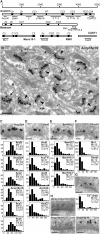
Fig. 1.
Fig. 1.
Stratified presynaptic localizations of sequence regions of Aczonin, Bassoon, Munc13, and RIM in conventional mammalian synapses. (A) Domain architectures of Aczonin, Bassoon, Munc13-1, RIM1, and CAST1, and sequence positions of the immunogens used in this study (Table 1). (B) Typical overview of preembedding silver-enhanced immunogold EM in the molecular layer of the rat cerebellum with Aczonin antibody anti-Aczp18p19 shows narrow bands of immunodecoration parallel to presynaptic PMs. Synapses sectioned vertically to the PM are marked by arrowheads. Synapses sectioned obliquely or almost parallel to the PM (arrows) can also be seen. (Scale bar: 500 nm.) (C) Six regions within the N-terminal 70% of the Aczonin sequence localize to distances of 57 to 79 nm from the presynaptic PM. Top: EM image representing this localization (anti-Aczp2p4). Column diagrams with the morphometric data of the six regions are shown below. For each dataset, the median PM distance in nm (“M”), the 95% CI in brackets, and the number of particles evaluated (“n”) are indicated. (Scale bars: 100 nm.) (D and E) Five regions within the C-terminal 30% of the Aczonin sequence and three regions in the C-terminal 10% of the Bassoon sequence localize to distances of 33 to 39 nm from the presynaptic PM. (F) The N-terminal 320 aa of Munc13-1/ubMunc13-2 (anti-Mu1), the N-terminal Rab3-binding sequences of RIM1/2α (anti-Rim43), and (G) a peptide sequence (aa 865–881) in the large central cytoplasmic loop of the P/Q-type Ca2+ channel α1A subunit localize to distances of 19 to 20 nm from the presynaptic PM. (H) Direct correlation between Aczonin localization and the physical dimensions of DPs. Sections of Lowicryl K4M-embedded samples subjected to adsorption staining with uranyl acetate/methyl cellulose displayed the regular array of DPs (Top Left). When these specimens were postembedding immunolabeled with anti-Aczp18p19 (Bottom Left; “post”), gold particles (arrows) were found right above the tips of DPs. The EM image (Bottom Right; “pre”) shows a synapse decorated with anti-Aczp18p19 by the preembedding technique as in BG. Note the negative membrane contrast in the Lowicryl-embedded samples.
Fig. 2.
Fig. 2.
Lateral topology of Aczonin and its interactor protein domains relative to DPs. DPs were visualized by EPTA staining, followed by preembedding immunogold labeling. (A) Four immunogens within the N-terminal 70% of the Aczonin sequence localize close to the DP axis. Top: Typical EM image (PM-vertical section; white arrowheads point to DPs). Column diagrams with the morphometric data are shown below. In the background of the diagrams, the lateral dimensions of DPs are indicated by a symbolic DP shape and three vertical dashed lines (DP center, 0 nm; width at base, ±28 nm). (B) Three immunogens within the C-terminal 30% of the Aczonin sequence localize to the DP periphery. (C) The Bassoon CT region and the Munc13 and RIM NT sequences localize to the DP periphery. For AC, Results includes information regarding determination and interpretation of the median and M* median values, and Table 2 provides a complete list of morphometric parameters. (D) A PM-parallel section of a specimen immunolabeled with anti-Rim43 illustrates that immunodecoration particles (arrows) lie at the sides of DPs. (E) An oblique section of an anti-Aczp18p19–labeled specimen illustrates that this immunogen lies above the DP layer. From bottom to top, one sees contiguous electron-dense material representing the postsynaptic density followed by spots representing DPs (arrowheads); as DPs expire further up, immunolabeling particles appear (arrows). (Scale bars: 100 nm.)
Fig. 3.
Fig. 3.
Differential localizations of Aczonin and CtBP2/Ribeye versus Bassoon, CAST1, Munc13, and RIM at photoreceptor ribbon synapses. (A) Aczonin (anti-Aczp18p19) is detected over the whole surface of ribbons, in vertical (Left) and horizontal sections (Middle), and of ribbon precursor spheres (Right). (B) Antibodies against nine Aczonin sequence regions and against CtBP2/Ribeye produce indiscriminable labeling patterns of the entire ribbon height. Left: Organization of ribbon synapse illustrated schematically, aligned to 10 column diagrams that show the morphometric evaluation of the vertical distributions of immunolabel for the different Aczonin regions and CtBP2/Ribeye. Distribution diagrams show the distances of particles from the PM ridge, normalized to the distance between the PM ridge (ordinate position, 0) and the ribbon tip (ordinate position, 1) of the respective synapse. The mean ordinate position of the ribbon base was 0.23. Right: Two representative EM images are shown. (C) The CT region of Bassoon, the NT sequence of RIM1/2α, the NT region of Munc13, and the central region of CAST1 localize to the ribbon base and/or the vesicle release site. Column diagrams for the vertical distributions of the four immunogens, and three representative EM images are shown. (Scale bars: 100 nm.) Table S1 provides a complete list of the morphometric and statistical parameters of the vertical and horizontal distributions.
Fig. 4.
Fig. 4.
Models for molecular topologies at DPs and the presynaptic particle grid. (A) Localizations of Aczonin, Bassoon, RIM, Munc13, and Cav2.1 calcium channel sequences are indicated in relation to the dimensions of DPs, synaptic vesicles (SV), and the PM as viewed from the side. Vertical and lateral localizations are combined from the results of Figs. 1 and 2. Approximate confidence intervals for the localizations, indicated by circular areas around the median values shown as crosses, are uniformly given as 10 nm for simplicity whereas the actual 95% CIs vary between 3 nm and 16 nm in the vertical direction and between 3 nm and 8 nm in the horizontal direction. White horizontal bars show localizations of immunogens for which only the vertical position could be determined; for such Aczonin sequences, the horizontal placements were interpolated from those of neighboring sequences, whereas the horizontal placement of the Ca2+ channel epitope is arbitrary. The approximate outline of the Aczonin protein in situ is indicated by a dark green silhouette of 20 nm horizontal diameter. This accounts for possible deviations of unmapped sequence intervals, and for finite true lateral localizations of the DP axis-near immunogens (presumably ≤5 nm but depicted here as zero for simplicity), which are obscured by the inaccuracy of the detection technique of approximately ±10 nm. (B) Model of a possible topology of Aczonin within the presynaptic particle grid. An idealized grid is shown as viewed from above, with DPs of 56 nm diameter at base and 72 nm center-to-center distance, and docked synaptic vesicles of 38 nm diameter (values determined in this study). As in A, Aczonin molecules are depicted as dimers centered on DPs. (Scale bar: 70 nm as the typical thickness of an ultrathin section for EM.)
Fig. P1.
Fig. P1.
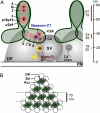
Immunogold EM determination of presynaptic protein topologies. (A) A synaptic terminal (ST), probably of the cholinergic splanchnic or vagus nerve and densely filled with neurotransmitter vesicles, contacts an adrenaline-secreting chromaffin cell (CAC) of the adrenal medulla. Several synaptic junctions are discernible by the higher protein densities particularly at the postsynaptic plasma membranes (PSD). Opposite them on the presynaptic side, one sees silver particles, appearing as black dots, which represent immunolabeling of a specific Aczonin sequence (p18p19, amino acids 1808–2150), forming a stripe at a defined distance from the plasma membrane. (BD) Simultaneous heavy metal staining of dense projections (white arrowheads) and immunostaining of protein segments (black dots) shows that Aczonin-p18p19 localizes more plasma membrane-distal and above dense projection tips whereas the C-terminal Aczonin-C2A segment (amino acids 4566–4744) and an N-terminal Munc13 sequence localize closer to the plasma membrane and between dense projections. (E) Model of molecular localizations and size proportions relative to dense projections (DP), synaptic vesicles (SV), and the presynaptic plasma membrane (PM). Aczonin “trunk” segments N, 2, 3, 4, p2p4, and p18p19 are within the N-terminal 70% of the protein sequence, whereas the “arm” segments 6, 7–6, 8, C2, and C2A are within the C-terminal 30% of Aczonin. Aczonin is hypothetically depicted as a dimer. (Scale bars: A, 500 nm; BD, 100 nm.)

References

    1. Wojcik SM, Brose N. Regulation of membrane fusion in synaptic excitation-secretion coupling: speed and accuracy matter. Neuron. 2007;55:11–24. - PubMed
    1. Schoch S, Gundelfinger ED. Molecular organization of the presynaptic active zone. Cell Tissue Res. 2006;326:379–391. - PubMed
    1. Phillips GR, et al. The presynaptic particle web: Ultrastructure, composition, dissolution, and reconstitution. Neuron. 2001;32:63–77. - PubMed
    1. Zhai RG, Bellen HJ. The architecture of the active zone in the presynaptic nerve terminal. Physiology (Bethesda) 2004;19:262–270. - PubMed
    1. Wang X, et al. A protein interaction node at the neurotransmitter release site: Domains of Aczonin/Piccolo, Bassoon, CAST, and RIM converge on the N-terminal domain of Munc13-1. J Neurosci. 2009;29:12584–12596. - PMC - PubMed

Publication types

MeSH terms

LinkOut - more resources