. 2011 Jul 19;108(29):11854-9.
doi: 10.1073/pnas.1107844108. Epub 2011 Jun 28.

Diverse two-cysteine photocycles in phytochromes and cyanobacteriochromes

Affiliations

Diverse two-cysteine photocycles in phytochromes and cyanobacteriochromes

Nathan C Rockwell et al. Proc Natl Acad Sci U S A. .

Abstract

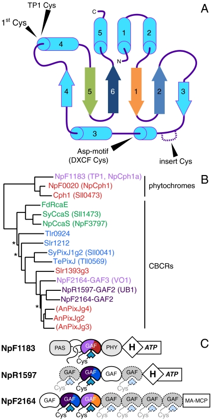
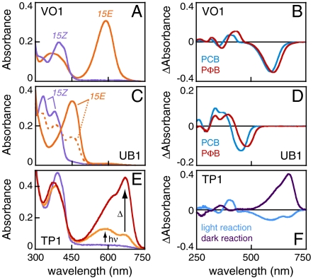
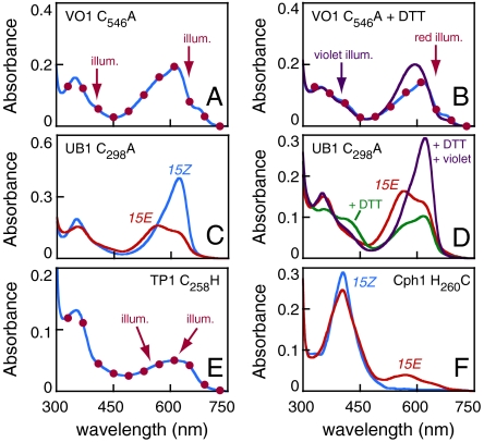
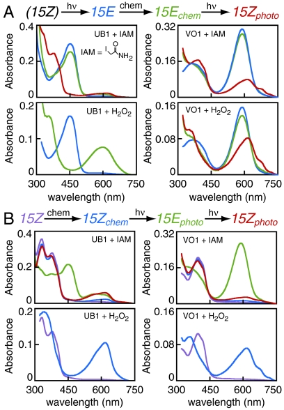
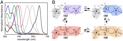
Phytochromes are well-known as photoactive red- and near IR-absorbing chromoproteins with cysteine-linked linear tetrapyrrole (bilin) prosthetic groups. Phytochrome photoswitching regulates adaptive responses to light in both photosynthetic and nonphotosynthetic organisms. Exclusively found in cyanobacteria, the related cyanobacteriochrome (CBCR) sensors extend the photosensory range of the phytochrome superfamily to shorter wavelengths of visible light. Blue/green light sensing by a well-studied subfamily of CBCRs proceeds via a photolabile thioether linkage to a second cysteine fully conserved in this subfamily. In the present study, we show that dual-cysteine photosensors have repeatedly evolved in cyanobacteria via insertion of a second cysteine at different positions within the bilin-binding GAF domain (cGMP-specific phosphodiesterases, cyanobacterial adenylate cyclases, and formate hydrogen lyase transcription activator FhlA) shared by CBCRs and phytochromes. Such sensors exhibit a diverse range of photocycles, yet all share ground-state absorbance of near-UV to blue light and a common mechanism of light perception: reversible photoisomerization of the bilin 15,16 double bond. Using site-directed mutagenesis, chemical modification and spectroscopy to characterize novel dual-cysteine photosensors from the cyanobacterium Nostoc punctiforme ATCC 29133, we establish that this spectral diversity can be tuned by varying the light-dependent stability of the second thioether linkage. We also show that such behavior can be engineered into the conventional phytochrome Cph1 from Synechocystis sp. PCC6803. Dual-cysteine photosensors thus allow the phytochrome superfamily in cyanobacteria to sense the full solar spectrum at the earth surface from near infrared to near ultraviolet.


Conflict of interest statement

The authors declare no conflict of interest.

Figures

Fig. 1.
Fig. 1.
Evolution of the photosensory GAF domain of phytochromes and CBCRs. (A) A topology diagram of the GAF domain, with the conserved “first Cys” and the three types of “second Cys” indicated. The first, second, fifth, and sixth beta strands are color-coded to match the sequence alignment in Fig. S3. (B) A neighbor-joining tree is shown for the GAF domains of published CBCRs and proteins described in this study, with names color-coded by ground-state absorbance. Branches with bootstrap values below 67% are indicated with an asterisk. Domains in parentheses bind bilin, but are photochemically inert (25). (C) Domain cartoons are shown for the proteins described in this work. CBCRs are color-coded by their photocycles, with near-UV absorbance represented by deep purple. Dashed GAF domains contain the first Cys, but have not yet been described.
Fig. 2.
Fig. 2.
Three newly identified dual-Cys biliprotein photosensors from Nostoc punctiforme. (A) Absorbance spectra are shown for VO1 with PCB. (B) Photochemical difference spectra are shown for VO1 with PCB (blue) and PΦB (red) chromophores. (C) Absorbance spectra are shown for UB1 with PCB. The photoequilibrium spectrum produced by 400 nm illumination is dashed orange, whereas that produced by 334 nm illumination is solid orange. (D) Photochemical difference spectra are shown for UB1 with PCB (blue) and PΦB (red) chromophores. (E) Absorbance spectra are shown for TP1 in the dark-adapted ground state (purple), after illumination with 400 ± 35 nm light (orange), and after subsequent incubation in darkness at 25 °C for 20 min (red). (F) Difference spectra are shown for photoconversion of TP1 with 400 nm light (blue) and for the subsequent thermal evolution of the 15E photoproduct (deep purple).
Fig. 3.
Fig. 3.
Identification of novel second Cys residues. (A) Absorbance spectra of C546A VO1. Illumination of the ground state (blue trace) with red light (650 ± 20 nm) or violet light (400 ± 35 nm) light produced indistinguishable spectra (red circles). (B) C546A VO1 supplemented with 50 mM DTT in the ground state (blue trace) and after illumination with saturating red light (red circles) or violet light (purple trace). (C) C298A UB1 in the 15Z (blue) and 15E (red) states. (D) C298A UB1 is shown in the 15E state before (red) and after (green) addition of 50 mM DTT. Illumination with violet light after addition of DTT regenerated the 15Z state (purple). (E) Absorbance spectra of C258H TP1. Illumination of the ground state (blue) with 600 ± 20 nm or 550 ± 35 nm light resulted in indistinguishable spectra (red circles). (F) Absorbance spectra are shown for H260C Cph1 in the ground state (blue) and after illumination with saturating 400 ± 35 nm light (red).
Fig. 4.
Fig. 4.
Characterization of the second linkage in VO1 and UB1. (A) Reverse photoconversion (15E to 15Z) was examined after treatment with IAM or H2O2. Photoproteins were converted to the 15E state (blue), followed by reaction with IAM or H2O2 in darkness to yield 15E chemical products (green). The 15E chemical products were then illuminated when appropriate to generate 15Z photoproducts (red). Blue traces for H2O2 treatment are scaled to reflect the 1∶1 dilution of sample upon peroxide addition. (B) Forward photoconversion was similarly examined. The 15Z photoproteins (purple) were treated with IAM or H2O2 in darkness to yield 15Z chemical products (blue), which were then cycled to the 15E state (green) and back (red).
Fig. 5.
Fig. 5.
Dual-cysteine photosensors. (A) Spectral coverage of representative dual-Cys photosensors. Absorbance spectra are shown for UB1 (blue), VO1 (green), and Tlr0924 [red (27)] in the 15Z (solid) and 15E (dashed) states. Wild-type 15Z Cph1 is shown in black for comparison. (B) A general dual-Cys photocycle. In the dark-adapted 15Z ground state (Upper Left), there is a covalent linkage between C10 and the second Cys. Light induces 15/16 photoisomerization to produce a 15E species with an intact second linkage (Upper Right). In UB1, this species is stable. In VO1, the second linkage is labile in the 15E state, red-shifting the photoproduct absorbance (Lower Right). Photoconversion produces a red-shifted species (bottom left). Only the B-, C-, and D-rings are shown; isomerization of the A-ring allows further spectral tuning.

References

    1. Montgomery BL, Lagarias JC. Phytochrome ancestry. Sensors of bilins and light. Trends Plant Sci. 2002;7:357–366. - PubMed
    1. Rockwell NC, Su YS, Lagarias JC. Phytochrome structure and signaling mechanisms. Annu Rev Plant Biol. 2006;57:837–858. - PMC - PubMed
    1. Auldridge ME, Forest KT. Bacterial phytochromes: More than meets the light. Crit Rev Biochem Mol Biol. 2011;46:67–88. - PubMed
    1. Franklin KA, Quail PH. Phytochrome functions in Arabidopsis development. J Exp Bot. 2010;61:11–24. - PMC - PubMed
    1. Giraud E, et al. Bacteriophytochrome controls photosystem synthesis in anoxygenic bacteria. Nature. 2002;417:202–205. - PubMed

Publication types

LinkOut - more resources