. 2011 Oct;96(10):1424-32.
doi: 10.3324/haematol.2011.042952. Epub 2011 Jun 28.

Over-expression of mitochondrial ferritin affects the JAK2/STAT5 pathway in K562 cells and causes mitochondrial iron accumulation

Affiliations

Over-expression of mitochondrial ferritin affects the JAK2/STAT5 pathway in K562 cells and causes mitochondrial iron accumulation

Paolo Santambrogio et al. Haematologica. 2011 Oct.

Abstract

Background: Mitochondrial ferritin is a nuclear encoded iron-storage protein localized in mitochondria. It has anti-oxidant properties related to its ferroxidase activity, and it is able to sequester iron avidly into the organelle. The protein has a tissue-specific pattern of expression and is also highly expressed in sideroblasts of patients affected by hereditary sideroblastic anemia and by refractory anemia with ringed sideroblasts. The present study examined whether mitochondrial ferritin has a role in the pathogenesis of these diseases.

Design and methods: We analyzed the effect of mitochondrial ferritin over-expression on the JAK2/STAT5 pathway, on iron metabolism and on heme synthesis in erythroleukemic cell lines. Furthermore its effect on apoptosis was evaluated on human erythroid progenitors.

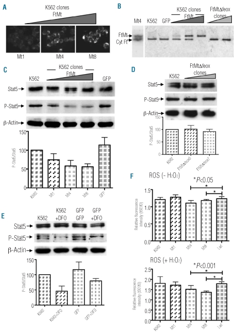
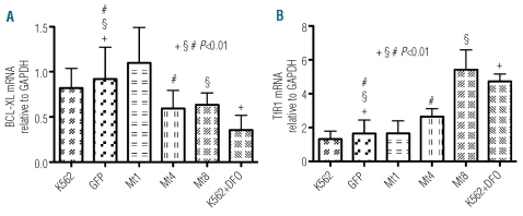
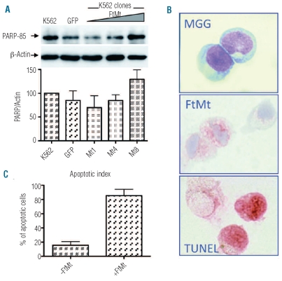
Results: Data revealed that a high level of mitochondrial ferritin reduced reactive oxygen species and Stat5 phosphorylation while promoting mitochondrial iron loading and cytosolic iron starvation. The decline of Stat5 phosphorylation induced a decrease of the level of anti-apoptotic Bcl-xL transcript compared to that in control cells; however, transferrin receptor 1 transcript increased due to the activation of the iron responsive element/iron regulatory protein machinery. Also, high expression of mitochondrial ferritin increased apoptosis, limited heme synthesis and promoted the formation of Perls-positive granules, identified by electron microscopy as iron granules in mitochondria.

Conclusions: Our results provide evidence suggesting that Stat5-dependent transcriptional regulation is displaced by strong cytosolic iron starvation status induced by mitochondrial ferritin. The protein interferes with JAK2/STAT5 pathways and with the mechanism of mitochondrial iron accumulation.


Figures

Figure 1.
Figure 1.
Effect of FtMt expression on K562 clones. (A) Immunostaining of the three selected clones (Mt1, Mt4 and Mt8) with the antibody specific for human FtMt. (B) Control and transduced K562 cells were grown in the presence of 1 μM 55Fe(III)-citrate and 10 μM ascorbic acid for 18 h. Soluble lysates were separated on 7% non-denaturing PAGE, dried and exposed to autoradiography. Positions of immunodection of Mt4 clone, probed with the specific anti-mitochondrial and anti-cytosolic ferritin antibodies by western blotting, and iron associated with mitochondrial (FtMt) and cytosolic (Cyt Ft) ferritins are indicated by the arrows. One representative experiment of three independent experiments is shown. (C) Stable clones and control K562 soluble cell lysates (25 μg per lane) were separated by 7.5% SDS-PAGE, blotted onto a nitrocellulose filter and probed with antibodies specific for total Stat5, phosphorylated Stat5 (P-Stat5) and β-actin. Arrows indicate positions of protein bands. Plots show band intensities of P-Stat5 relative to total Stat5 representative of three independent experiments. Triangles indicate increasing amount of expressed protein. (D) Stable FtMtΔfeox clones and control K562 soluble cell lysates were analyzed as in panel (C). (E) Control, GFP transduced and DFO-treated K562 cells were analyzed as in panel (C). (F) Control and transduced K562 cells were loaded with a ROS-sensitive fluorescent probe DHR123 and treated (+ H2O2) or not (− H2O2) with H2O2 for 30 min. Plots represent mean (±SD) of ROS production after 30 min of incubation with or without H2O2 treatment (t30′) relative to basal ROS level (t0). Three independent experiments, were performed each in octuplicate. Significant changes are marked.
Figure 2.
Figure 2.
Effect of FtMt expression on transcription of Stat5 target genes in K562 clones. Control, transfected and DFO-treated K562 cells were harvested for isolation of total RNA. Levels of mRNA expression were determined by quantitative real-time PCR for Bcl-xL (A) and TfR1 (B) and then normalized to mRNA expression of GAPDH. Results are presented as mRNA amount relative to GAPDH (±SD). Four independent experiments were performed each in triplicate. Significant changes compared to GFP control cells are marked.
Figure 3.
Figure 3.
Effect of FtMt expression on apoptosis in K562 and CD34+ cells. (A) Stable clones and control K562 were treated with DFO, soluble cell lysates (25 μg per lane) were separated by 10% SDS PAGE, blotted onto a nitrocellulose filter and probed with antibodies specific for the p85 PARP fragment (PARP-85) and β-actin. Plots show band intensities of the PARP fragment relative to β-actin. Results are representative of three independent experiments. Arrows indicate positions of protein bands. Triangles indicate increasing amount of expressed protein. (B) CD34+ bone marrow cells were isolated from normal donors, transduced with LV-FtMt and cultured for 21 days. After 14 and 21 days, cells were analyzed for morphology by May Grünwald Giemsa staining (MGG), for FtMt expression by immunocytochemistry (FtMt) and for apoptosis by a TUNEL assay (TUNEL). Representative samples at day 14. (C) Plots represent the apoptotic index of FtMt expressing (+FtMt) and non-expressing (−FtMt) cells, evaluated by a double immunocytochemistry technique, at day 14.
Figure 4.
Figure 4.
Effect of FtMt expression on mitochondrial iron load and heme synthesis. (A) K562 Mt8 and MtΔfeox7 clones were grown for 6 days in the presence of 20 μM Fe-Tf and then stained for iron content with Perls’ reaction (left panels) or immunostained with an antibody specific for FtMt (right panels). The arrows point to granules positive for iron staining. (B) K562 cells and clones treated as in (A) were analyzed by electron microscopy. Upper panels show the controls, MtΔfeox7 and K562 cells, stained with uranyl and lead citrate. The second row shows a portion of Mt8 cells with a group of mitochondria stained with uranyl and lead citrate (left panel). The area selected in the white square is enlarged in the right panel, the arrows point to electrondense spots. The panels in the third row show the presence of granules in unstained Mt8 cells; the square selected in white is enlarged in the right panel. The bottom panels show the conventional ultrastructural organization (left panel) and the iron map (red color) superimposed on the ultra-structural organization of the same field obtained at 250 eV (right panel) obtained by ESI (see Design and Methods section). Bars indicate 500 nm. (C) Control and transfected MEL cells were grown for 3 days in the presence of 1.5% DMSO, and heme production was evaluated by a chemical method. Plots represent mean (±SD) of heme content relative to DMSO-treated control cells. Three independent experiments, were performed each in duplicate. Significant changes are marked.

References

    1. Levi S, Corsi B, Bosisio M, Invernizzi R, Volz A, Sanford D, et al. A human mitochondrial ferritin encoded by an intronless gene. J Biol Chem. 2001;276(27):24437–40. - PubMed
    1. Arosio P, Levi S. Cytosolic and mitochondrial ferritins in the regulation of cellular iron homeostasis and oxidative damage. Biochim Biophys Acta. 2010;1800(8):783–92. - PubMed
    1. Drysdale J, Arosio P, Invernizzi R, Cazzola M, Volz A, Corsi B, et al. Mitochondrial ferritin: a new player in iron metabolism. Blood Cells Mol Dis. 2002;29(3):376–83. - PubMed
    1. Langlois d'Estaintot B, Santambrogio P, Granier T, Gallois B, Chevalier JM, Precigoux G, et al. Crystal structure and biochemical properties of the human mitochondrial ferritin and its mutant Ser144Ala. J Mol Biol. 2004;340(2):277–93. - PubMed
    1. Bou-Abdallah F, Santambrogio P, Levi S, Arosio P, Chasteen ND. Unique iron binding and oxidation properties of human mitochondrial ferritin: a comparative analysis with human H-chain ferritin. J Mol Biol. 2005;347(3):543–54. - PubMed

Publication types

MeSH terms