. 2011 Jun 28;30(15):3173-85.
doi: 10.1038/emboj.2011.215.

A novel strategy for evasion of NK cell immunity by tumours expressing core2 O-glycans

Affiliations

A novel strategy for evasion of NK cell immunity by tumours expressing core2 O-glycans

Shigeru Tsuboi et al. EMBO J. .

Abstract

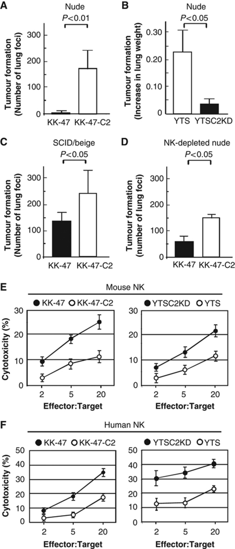
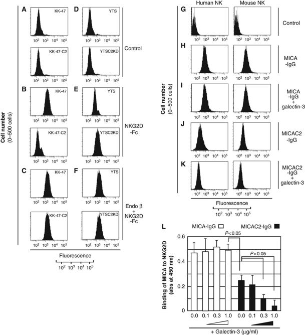
The O-glycan branching enzyme, core2 β-1,6-N-acetylglucosaminyltransferase (C2GnT), forms O-glycans containing an N-acetylglucosamine branch connected to N-acetylgalactosamine (core2 O-glycans) on cell-surface glycoproteins. Here, we report that upregulation of C2GnT is closely correlated with progression of bladder tumours and that C2GnT-expressing bladder tumours use a novel strategy to increase their metastatic potential. Our results showed that C2GnT-expressing bladder tumour cells are highly metastatic due to their high ability to evade NK cell immunity and revealed the molecular mechanism of the immune evasion by C2GnT expression. Engagement of an NK-activating receptor, NKG2D, by its tumour-associated ligand, Major histocompatibility complex class I-related chain A (MICA), is critical to tumour rejection by NK cells. In C2GnT-expressing bladder tumour cells, poly-N-acetyllactosamine was present on core2 O-glycans on MICA, and galectin-3 bound the NKG2D-binding site of MICA through this poly-N-acetyllactosamine. Galectin-3 reduced the affinity of MICA for NKG2D, thereby severely impairing NK cell activation and silencing the NK cells. This new mode of NK cell silencing promotes immune evasion of C2GnT-expressing bladder tumour cells, resulting in tumour metastasis.


Conflict of interest statement

The authors declare that they have no conflict of interest.

Figures

Figure 1
Figure 1
Expression of core2 O-glycans in bladder tumours. (A) Biosynthesis pathway of mucin-type O-glycan core structures, core1–4. N-acetylgalactosamine (GalNAc); N-acetylglucosamine (GlcNAc); Galactose (Gal). GalNAc is transferred to serine (Ser) or threonine (Thr) residues in a polypeptide by GalNAc-T, peptide GalNAc transferase. GalNAcα1-Ser/Thr is converted by Core1 synthase to Galβ1-3GalNAcα1-Ser/Thr (core1). Core1 is then converted by C2GnT to core2. Core1 is also converted by Core3 synthase to core3. Core3 is converted by C2GnT-2 to core4. Only C2GnT-2 is capable to synthesize core4 (Lee et al, 2009). (B) Immunohistochemistry of bladder tumours using anti-C2GnT antibody. C2GnT-positive tumour (upper panel); C2GnT-negative tumour (lower panel). C2GnT staining of a C2GnT-positive tumour specimen exhibited cytoplasmic pattern close to the nucleus. Two typical C2GnT-positive cells are denoted by arrows (upper panel). Bar, 10 μm. (C) Biosynthesis of core2 O-glycans in C2GnT-positive tumours. Oligosaccharide analysis was carried out on C2GnT-positive and C2GnT-negative tumours. 3H-labelled O-glycans were isolated, desialylated and subjected to Bio-Gel P4 (1.0 × 100 cm) gel filtration. The elution profiles obtained from C2GnT-positive tumours (upper panel) and C2GnT-negative tumours (lower panel) were shown here. The elution position of a core2 branch containing tetrasaccharide, Galβ1-3(Galβ1−4GlcNAcβ1-6)GalNAcOH was indicated by an arrow. (D) Kaplan–Meier curve for the cause-specific survival of bladder tumour patients according to C2GnT staining status. Fifty-seven bladder tumour patients were divided into two groups according to the expression status of C2GnT, C2GnT positive (n=37, red line) and C2GnT negative (n=20, blue line).
Figure 2
Figure 2
Evasion from NK cell tumour rejection responses by C2GnT-expressing tumour cells. (A, B) Tumour formation by bladder tumour cells in nude mice. Bladder tumour cells were intravenously injected into nude mice. After 3 weeks, the lungs were examined for tumour formation by counting metastatic foci or measuring lung weights. KK-47, closed bar (n=8); KK-47-C2, open bar (n=6) (A). YTS, open bar (n=7); YTSC2KD, closed bar (n=7) (B). (C, D) Tumour formation by bladder tumour cells in mice with impaired NK cell cytotoxicity. Bladder tumour cells were intravenously injected into SCID/beige mice (n=5 for each cell) (C) and NK cell-depleted nude mice (n=5 for each cell) (D). After 3 weeks, the lungs were examined for tumour formation by counting metastatic foci. KK-47, closed bars and KK-47-C2, open bars. (E, F) Effect of C2GnT expression on NK cell cytotoxicity. Cytotoxicity of mouse NK cells (E) and human NK cells (F) against bladder tumour cells was assayed. Mean values±s.e. of three independent experiments.
Figure 3
Figure 3
C2GnT-expressing tumour cells express MICA carrying poly-N-acetyllactosamine. (A) Biosynthesis of poly-N-acetyllactosamine on O-glycans. β-1,4-Galactosyltransferase IV (β1-4Gal-T IV) together with β-1,3-N-acetylglucosaminyltransferase (β1-3 GlcNAc-T) synthesize poly-N-acetyllactosamine in core2 branched oligosaccharides. Lycoperiscon esculentum (tomato) lectin (LEL) and galectin-3 bind specifically to poly-N-acetyllactosamine with at least three lactosamine unit repeats. (B, C) MICA from KK-47-C2 (B) and YTS (C) carries poly-N-acetyllactosamine. Total lysates from benzyl 2-acetamido-2-deoxy-α-D-galactopyranoside (BAG, 2 mM)-treated or untreated cells (5 × 10⁶ cells) were analysed by western blotting with anti-MCA (clone BAMO1) (lanes 1–4). Total lysates from tunicamycin (0.5 μg/ml)-treated cells were immunoprecipitated with LEL-agarose followed by western blot analysis with anti-MICA (lanes 5 and 6).
Figure 4
Figure 4
MICA and galectin-3 on the surface of bladder tumour cells. (AJ) Flow cytometric analyses of bladder tumour cells. Cells were stained with control IgG2a (A, F) and anti-MICA (clone BAMO3) (B, G). Tunicamycin-treated cells were stained with IgG1 (C, H) and anti-galectin-3 (D, I). Tunicamycin-treated cells (1 × 10⁶) were incubated with endo-β-galactosidase (80 mU/ml, 37°C, 8 h), and then stained with anti-galectin-3 (E, J). (K, L) Galectin-3 binds MICAC2. Cells were subjected to dithio-bis-sulphosuccinimydyl propionate (DTSSP) crosslinking of cell-surface proteins and the total lysates were immunoprecipitated with anti-MICA (clone BAMO3) followed by western blotting with anti-MICA (clone BAMO1) (lanes 1–4) and anti-galectin-3 (lanes 5–8). Galectin-3 co-immunoprecipitated with MICAC2 from C2GnT-expressing KK-47-C2 (lane 6) (K) and YTS cells (lane 5) (L). Binding of galectin-3 to MICAC2 was disrupted by endo-β-galactosidase treatment (lanes 7 and 8) (K, L).
Figure 5
Figure 5
Galectin-3 binds MICAC2 from C2GnT-positive bladder tumour specimens. (A) MICA from C2GnT-positive bladder tumour specimens carries poly-N-acetyllactosamine. Total lysates from patients' tumour specimens (P1, P2, P3, and P4) were analysed by western blotting with anti-MICA (clone BAMO1) (lanes 1–4). The MICA immunoprecipitates from tumour specimens (0.1 g) with anti-MICA (clone BAMO3) were treated with Glycopeptidase F (20 μU/μl, 37°C, 16 h) and then re-immunoprecipitated with LEL-agarose followed by western blotting with anti-MICA (clone BAMO1) (lanes 5–8). (B) Bladder tumour specimens from patients (P1, P2, P3, and P4) were subjected to DTSSP crosslinking of cell-surface proteins and the total lysates were analysed by western blotting with anti-galectin-3 (lanes 1–4). The MICA immunoprecipitates from tumour specimens with anti-MICA (clone BAMO3) were treated with Glycopeptidase F and then analysed by western blotting with anti-MICA (BAMO1) (lanes 5–8) and anti-galectin-3 (lanes 9–12).
Figure 6
Figure 6
Galectin-3 binding to MICA reduces the interaction of NKG2D with MICA. (AH) Interaction of NKG2D with MICA on the surface of bladder tumour cells. Flow cytometric analysis of bladder tumour cells (AF) and NK cells (G, H). Bladder tumour cells were stained with control IgG chimera (CD43-IgG) (A, D) and NKG2D-Fc (B, E). Cells were treated with endo-β-galactosidase followed by NKG2D-Fc staining (C). (GK) Human (left panel) and mouse (right panel) NK cells were stained with CD43-IgG, MICA-IgG, or MICAC2-IgG. (L) Effect of galectin-3 on MICA binding to NKG2D. MICA-IgG or MICAC2-IgG was incubated with galectin-3 at 37°C for 30 min, and then added to the plate coated with NKG2D-Fc by ELISA (MICA-IgG, open bars; MICAC2-IgG, closed bars). Mean values±s.e. of three independent experiments.
Figure 7
Figure 7
Galectin-3 binds MICAC2, impairing NK cell functions. (A, B) Effect of galectin-3 on secretion of INF-γ (A) and granzyme B (B). NK cells were stimulated with the plates coated with CD43-IgG (cntl.), MICA-IgG, or MICAC2-IgG in the presence (1.0 μg/ml) and absence of galectin-3. After stimulation, the supernatant was recovered and NK cell activation was evaluated by measuring secretion of INF-γ and granzyme B into the supernatant. (C, D) Removal of galectin-3 from bladder tumour cells enhances NK-mediated cytotoxicity. Tunicamycin-treated bladder tumour cells were treated with endo-β-galactosidase, and then human NK-mediated cytotoxicity was measured at an effector:target ratio of 5:1. Mean values±s.e. of three independent experiments.
Figure 8
Figure 8
In vivo role of NKG2D and tumour cell-surface-bound galectin-3 in rejection responses of the bladder tumours. (A) Mouse NK cells were analysed for cytotoxicity against bladder tumour cells in the presence of indicated antibodies at an effector:target ration of 5:1. Mouse NK cells were pre-incubated for 30 min with saturating concentration of rat IgG1 (+cntl.) or anti-mouse NKG2D blocking antibody (CX5). (B) Bladder tumour cells (5 × 10⁶ cells) were intravenously injected into nude mice. The nude mice were pre-treated with anti-mouse NKG2D (CX5) (n=5 for each cell) or control rat IgG1 (+cntl.) (n=3 for each cell). After 3 weeks, the lungs were examined for tumour formation by counting metastatic foci. (C) Mouse NK cells were assayed for cytotoxicity against bladder tumour cells after endo-β-galactosidase treatment at an effector:target ratio of 5:1. Tumour cells were pre-treated with (+) (80 mU/ml, 37°C, 8 h) or without endo-β-galactosidase treatment (−). (D) Endo-β-galactosidase-treated bladder tumour cells (5 × 10⁶ cells) (KK-47, closed bars (n=4); KK-47-C2, open bars (n=4)) were intravenously injected into nude mice. After 3 weeks, the lungs were examined for tumour formation. The results from the tumour formation by untreated cells (Figure 2A) were re-displayed as a left panel of Figure 8D for the comparison purpose.

Similar articles

Cited by

References

    1. Abdou AG, Hammam MA, Farargy SE, Farag AG, El Shafey EN, Farouk S, Elnaidany NF (2010) Diagnostic and prognostic role of galectin 3 expression in cutaneous melanoma. Am J Dermatopathol 32: 809–814 - PubMed
    1. Argueso P, Guzman-Aranguez A, Mantelli F, Cao Z, Ricciuto J, Panjwani N (2009) Association of cell surface mucins with galectin-3 contributes to the ocular surface epithelial barrier. J Biol Chem 284: 23037–23045 - PMC - PubMed
    1. Bahram S, Bresnahan M, Geraghty DE, Spies T (1994) A second lineage of mammalian major histocompatibility complex class I genes. Proc Natl Acad Sci USA 91: 6259–6263 - PMC - PubMed
    1. Borsig L, Wong R, Hynes RO, Varki NM, Varki A (2002) Synergistic effects of L- and P-selectin in facilitating tumor metastasis can involve non-mucin ligands and implicate leukocytes as enhancers of metastasis. Proc Natl Acad Sci USA 99: 2193–2198 - PMC - PubMed
    1. Brunda MJ, Luistro L, Warrier RR, Wright RB, Hubbard BR, Murphy M, Wolf SF, Gately MK (1993) Antitumor and antimetastatic activity of interleukin 12 against murine tumors. J Exp Med 178: 1223–1230 - PMC - PubMed

Publication types

MeSH terms

Substances