. 2011 Dec;31(12):2302-12.
doi: 10.1038/jcbfm.2011.90. Epub 2011 Jun 29.

SK2 channels are neuroprotective for ischemia-induced neuronal cell death

Affiliations

SK2 channels are neuroprotective for ischemia-induced neuronal cell death

Duane Allen et al. J Cereb Blood Flow Metab. 2011 Dec.

Abstract

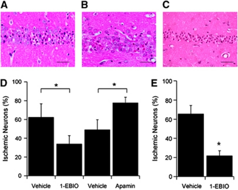
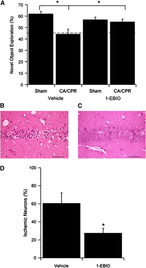
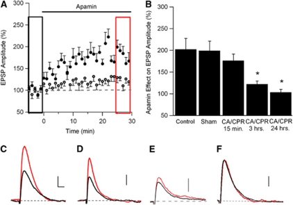
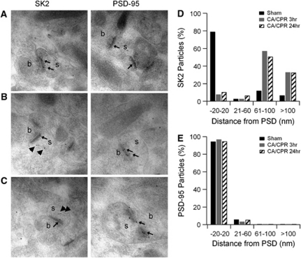
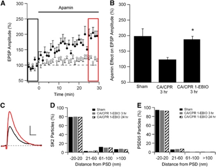
In mouse hippocampal CA1 pyramidal neurons, the activity of synaptic small-conductance Ca(2+)-activated K(+) channels type 2 (SK2 channels) provides a negative feedback on N-methyl-D-aspartate receptors (NMDARs), reestablishing Mg(2+) block that reduces Ca(2+) influx. The well-established role of NMDARs in ischemia-induced excitotoxicity led us to test the neuroprotective effect of modulating SK2 channel activity following cerebral ischemia induced by cardiac arrest and cardiopulmonary resuscitation (CA/CPR). Administration of the SK channel positive modulator, 1-ethyl-benzimidazolinone (1-EBIO), significantly reduced CA1 neuron cell death and improved CA/CPR-induced cognitive outcome. Electrophysiological recordings showed that CA/CPR-induced ischemia caused delayed and sustained reduction of synaptic SK channel activity, and immunoelectron microscopy showed that this is associated with internalization of synaptic SK2 channels, which was prevented by 1-EBIO treatment. These results suggest that increasing SK2 channel activity, or preventing ischemia-induced loss of synaptic SK2 channels, are promising and novel approaches to neuroprotection following cerebral ischemia.


Figures

Figure 1
Figure 1
Pharmacological enhancement of SK2 channel activity increases survival of CA1 neurons following cardiac arrest (CA). (A) Representative photomicrograph of hippocampal CA1 neurons from a wild-type (WT) mouse injected with vehicle (intraperitoneal injection) 30 minutes before and 6 hours after CA and cardiopulmonary resuscitation (CA/CPR) and stained with hematoxylin and eosin (H&E) 3 days later. Damaged neurons identified by the presence of pink eosinophilic cytoplasm and a dark pyknotic nucleus. (B) Representative photomicrograph of hippocampal CA1 neurons from a WT mouse injected with the SK2, Ca2+-sensitivity-enhancing drug 1-ethyl-benzimidazolinone (1-EBIO) (16 mg/kg, intraperitoneally), 30 minutes before and 6 hours after CA/CPR and stained with H&E 3 days later. Damaged neurons identified by the presence of pink eosinophilic cytoplasm and a dark pyknotic nucleus. (C) Representative photomicrograph of hippocampal CA1 neurons from a WT mouse injected with the SK2 antagonist apamin (0.1 mg/kg, intraperitoneally), 30 minutes before and 6 hours after CA/CPR and stained with H&E 3 days later. (D) Quantification of ischemic neurons in the CA1 region of the hippocampus 3 days after CA/CPR. (E) Quantification of ischemic neurons in the CA1 region of the hippocampus 3 days after CA/CPR in mice treated with 16 mg/kg 1-EBIO 30 minutes after resuscitation, with a second injection given 6 hours after resuscitation. Data are presented as mean±s.e.m. *P<0.05 compared with vehicle.
Figure 2
Figure 2
Pharmacological enhancement of SK channel activity improves long-term cognitive recovery. (A) Novel object recognition was assessed 7 days after cardiac arrest and cardiopulmonary resuscitation (CA/CPR) or sham-operated in vehicle or 1-ethyl-benzimidazolinone (1-EBIO) (16 mg/kg administered 30 minutes after resuscitation)-treated mice. The dashed line represents a preference ratio of 50%, indicating a lack of memory of the novel object. CA/CPR significantly reduced novel object preference in vehicle-treated mice, which was significantly higher in 1-EBIO-treated mice after CA/CPR. (B) Representative photomicrograph of hippocampal CA1 neurons from a wild-type (WT) mouse injected with vehicle 30 minutes after CA/CPR and stained with hematoxylin and eosin (H&E) 10 days later. Damaged neurons identified by the presence of pink eosinophilic cytoplasm and a dark pyknotic nucleus. (C) Representative photomicrograph of hippocampal CA1 neurons from a WT mouse injected with 1-EBIO (16 mg/kg, intraperitoneally), 30 minutes after after CA/CPR and stained with H&E 10 days later. Damaged neurons identified by the presence of pink eosinophilic cytoplasm and a dark pyknotic nucleus. (D) Quantification of ischemic neurons in the CA1 region of the hippocampus 10 days after CA/CPR in mice treated with 1-EBIO 30 minutes and again 6 hours after resuscitation. Data are presented as mean±s.e.m. *P<0.05 compared with vehicle.
Figure 3
Figure 3
Ischemia is associated with the loss of synaptic SK2 channel activity in CA1 neurons by 3 hours, but not by 15 minutes. (A) Time course of EPSP amplitude (mean±s.e.m.) from sham-operated mice (solid circles) and mice 3 hours after cardiac arrest and cardiopulmonary resuscitation (CA/CPR) (empty circles) before (black rectangle) and after (red rectangle) a 30-minute application of apamin (100 nmol/L). Apamin was applied at time 0. Each point is the average of two adjacent, normalized EPSPs. (B) Quantification of blocking synaptic SK2 channels with apamin on EPSP amplitude. Average EPSP amplitude (mean±s.e.m.) 30 minutes after adding apamin (100 nmol/L, red square in (A) normalized to baseline (black square in (A). *P<0.05 compared with sham controls. (C) Example EPSPs from sham-operated mice before (black) and after (red) a 30-minute application of apamin (100 nmol/L). All example EPSPs are the average of 15 EPSPs. (D) Example EPSPs from mice 15 minutes after CA/CPR before (black) and after (red) a 30-minute application of apamin (100 nmol/L). (E) Example EPSPs from mice 3 hours after CA/CPR before (black) and after (red) a 30-minute application of apamin (100 nmol/L). (F) Example EPSPs from mice 24 hours after CA/CPR before (black) and after (red) a 30-minute application of apamin (100 nmol/L). Scale bars on traces are vertical: 1 mV; horizontal: 50 milliseconds (scale same in all traces). The color reproduction of this figure is available at the Journal of Cerebral Blood Flow and Metabolism journal online. EPSP, excitatory post-synaptic potential.
Figure 4
Figure 4
SK2 channels are removed from the postsynaptic density following cardiac arrest and cardiopulmonary resuscitation (CA/CPR). (A) Single immunogold labeling for SK2 or postsynaptic density (PSD)-95 in representative asymmetric synapses of hippocampal CA1 neurons in sham control mice or 3 (B) or 24 hours (C) following CA/CPR. Small arrows indicate labeling near the PSD and bold arrows indicate SK2 immunogold particles that appear distant from the PSD in post-CA/CPR mice. (D) Quantification of immunogold particles for SK2 expressed as percentage of total particles in each of four distance bins from the PSD (in nm). Black bars are the sham control condition, gray bars 3 hours after CA/CPR, and stripped bars 24 hours after CA/CPR. (E) Quantification of immunogold particles for PSD-95 expressed as percentage of total particles in each of four distance bins from the PSD (in nm). Black bars are the control condition, gray bars 3 hours after CA/CPR, and stripped bars 24 hours after CA/CPR.
Figure 5
Figure 5
Injection with 1-ethyl-benzimidazolinone (1-EBIO) rescues the internalization of SK2 channels following cardiac arrest and cardiopulmonary resuscitation (CA/CPR). (A) Time course of EPSP amplitude (mean±s.e.m.) before (black rectangle) and after (red rectangle) a 30-minute application of apamin (100 nmol/L). Mice that received CA/CPR following an injection with 1-EBIO (solid circles) 30 minutes beforehand are compared with mice that received only CA/CPR (empty circles). Both groups are assessed 3 hours after CA/CPR. Apamin was applied at time 0 and each point is the average of two adjacent, normalized EPSPs. (B) Quantification of blocking synaptic SK2 channels with apamin on EPSP amplitude. Sham and CA/CPR 3 hours time point data from Figure 3 are replotted for better comparison with 1-EBIO preinjected 3 hours time point data. Average EPSP amplitude (mean±s.e.m.) 30 minutes after adding apamin (100 nmol/L, red square in Figures 3A or 5A) is normalized to baseline (black square in Figures 3A or 5A). *P<0.05 compared with CA/CPR 3 hours. (C) Example EPSPs from mice injected 30 minutes before CA/CPR with 1-EBIO and assessed 3 hours after resuscitation before (black) and after (red) a 30-minute application of apamin (100 nmol/L). All example EPSPs are the average of 15 EPSPs. (D) Quantification of immunogold particles for SK2 expressed as percentage of total particles in each of four distance bins from the postsynaptic density (PSD) (in nm). Black bars are the sham control condition, gray bars 3 hours after CA/CPR+1-EBIO, and stripped bars 24 hours after CA/CPR+1-EBIO. (E) Quantification of immunogold particles for PSD-95 expressed as percentage of total particles in each of four distance bins from the PSD (in nm). Black bars are the control condition, gray bars 3 hours after CA/CPR, and stripped bars 24 hours after CA/CPR. The color reproduction of this figure is available at the Journal of Cerebral Blood Flow and Metabolism journal online. EPSP, excitatory post-synaptic potential.

References

    1. Allen D, Bond CT, Lujan R, Ballesteros-Merino C, Lin MT, Wang K, Klett N, Watanabe M, Shigemoto R, Stackman RW, Maylie J, Adelman JP. The SK2-long isoform directs synaptic localization and function of SK2-containing channels. Nature Neuroscience. 2011;14:744–749. - PMC - PubMed
    1. Ballanyi K. Protective role of neuronal KATP channels in brain hypoxia. J Exp Biol. 2004;207:3201–3212. - PubMed
    1. Benveniste H, Drejer J, Schousboe A, Diemer NH. Elevation of the extracellular concentrations of glutamate and aspartate in rat hippocampus during transient cerebral ischemia monitored by intracerebral microdialysis. J Neurochem. 1984;43:1369–1374. - PubMed
    1. Bernard SA, Gray TW, Buist MD, Jones BM, Silvester W, Gutteridge G, Smith K. Treatment of comatose survivors of out-of-hospital cardiac arrest with induced hypothermia. N Engl J Med. 2002;346:557–563. - PubMed
    1. Boast CA, Gerhardt SC, Pastor G, Lehmann J, Etienne PE, Liebman JM. The N-methyl-D-aspartate antagonists CGS 19755 and CPP reduce ischemic brain damage in gerbils. Brain Res. 1988;442:345–348. - PubMed

Publication types

MeSH terms

Substances

LinkOut - more resources