

New Intraspinal cause of physiological FDG uptake
- PMID: 21712915
- PMCID: PMC3109827
- DOI: 10.4103/0972-3919.78257
New Intraspinal cause of physiological FDG uptake
Abstract
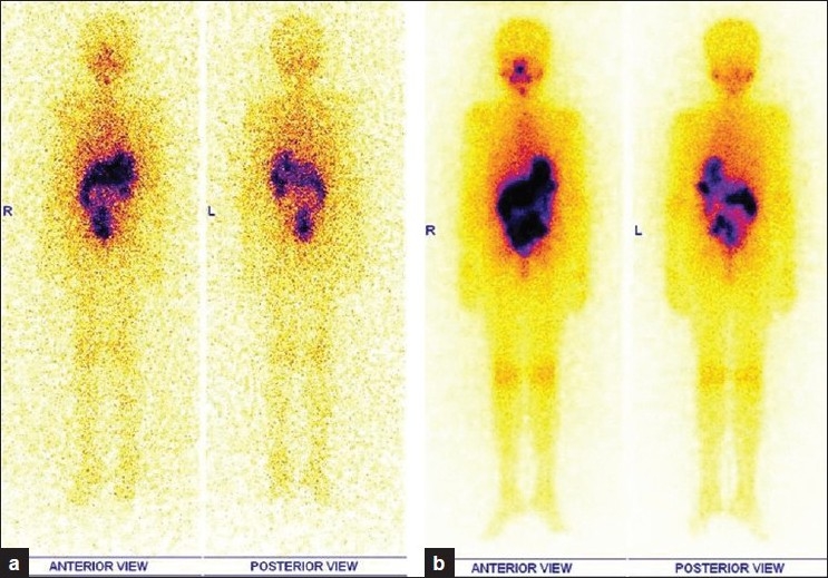
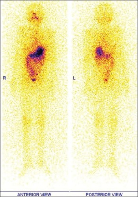
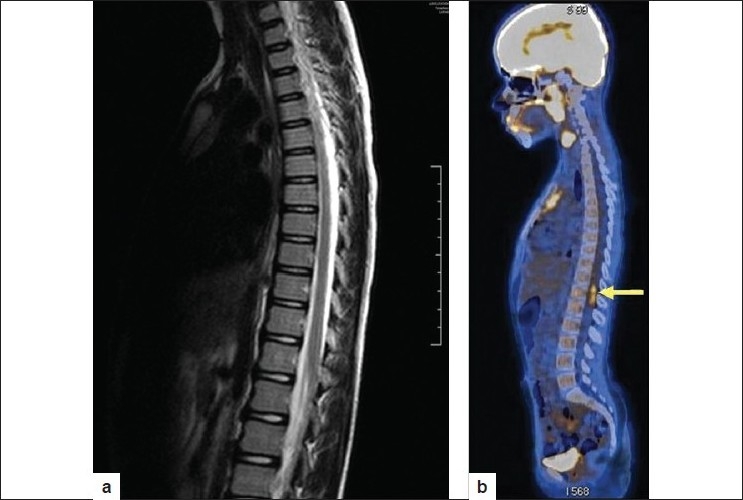
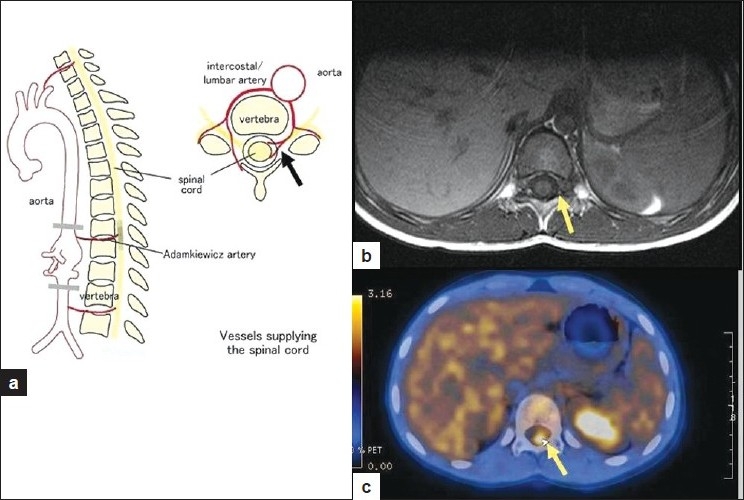
We present a paediatric case of Papillary Ca thyroid under evaluation for elevated Thyroglobulin (Tg) level with negative (131)I wholebody scintigraphy. Differentiated thyroid cancer (DTC) arises from follicular epithelium and retains basic biological features like expression of sodium iodide symporter (NIS), which is the cellular basis of radio iodine ((131)I) concentration during thyroid ablation. Once dedifferentiation of thyroid cells occurs, cells fail to concentrate (131)I, posing both diagnostic and therapeutic problems in DTC and one may have to resort to other imaging techniques for disease localization. As DTC progression is slow, patients have a relatively good prognosis. However children with thyroid malignancies need aggressive management, as initial presentation itself maybe with nodal metastases. It is well known that FDG PET CT apart from its oncological applications, is also used in the evaluation of vascular inflammation especially Takayasu's arteritis. It is also reported in literature, that (18)F-FDG uptake can be seen relatively frequently in the arterial tree of cancer patients. Dunphy et al reported the association of vascular FDG uptake in inflammation as well as in normal arteries. This study typically describes FDG uptake in a patchwork of normal vessel, focal inflammation and or calcification of vessels. The other plausible reasons for significant vascular (18)F-FDG uptake are drugs such as potent non steroidal anti-inflammatory agents, dexamethasone, prednisone and tacrolimus. Our patient showed false positive (18)F Fluorodeoxyglucose (FDG) uptake in spinal cord at D11/12 and D12/L1 vertebral levels in FDG PET CT imaging performed as part of raised Thyroglobulin workup. This intra spinal FDG uptake is attributed to physiological uptake and inadequate FDG clearance from artery of Adamkiewicz, which can be added as a new physiological cause of FDG uptake unreported in literature as yet.
Keywords: Artery of Adamkiewicz; Dedifferentiated thyroid cancer; FDG PET scan; papillary Ca thyroid; radioiodine I 131 scan; thyroglobulin.
Conflict of interest statement
Figures
References
-
- Schlüter B, Bohuslavizki KH, Beyer W, Plotkin M, Buchert R, Clausen M. Impact of FDG PET on patients with differentiated thyroid cancer who present with elevated thyroglobulin and negative 131I Scan. J Nucl Med. 2001;42:71–6. - PubMed
-
- Yun M, Jang S, Cucchiara A, Newberg AB, Alavi A. 18F FDG uptake in the large arteries: A correlation study with the atherogenic risk factors. Semin Nucl Med. 2002;32:70–6. - PubMed
-
- Tatsumi M, Cohade C, Nakamoto Y, Wahl RL. Fluorodeoxyglucose uptake in the aortic wall at PET/CT: Possible finding for active atherosclerosis. Radiology. 2003;229:831–7. - PubMed
-
- Dunphy MP, Freiman A, Larson SM, Strauss HW. Association of Vascular 18F-FDG Uptake with Vascular Calcification. J Nucl Med. 2005;46:1278–84. - PubMed
-
- Abdul Jalil N, Abdul Rahim N, Md Shalleh N, Rossetti C. 18F-FDG positron emission tomography/ computed tomography and the “underground map” appearance in imaging Horton’s arteritis. Singapore Med J. 2008;49:e178. - PubMed
Publication types
LinkOut - more resources
Full Text Sources
Miscellaneous
