. 2011;6(6):e21118.
doi: 10.1371/journal.pone.0021118. Epub 2011 Jun 21.

Serum soluble HLA-E in melanoma: a new potential immune-related marker in cancer

Affiliations

Serum soluble HLA-E in melanoma: a new potential immune-related marker in cancer

Mathilde Allard et al. PLoS One. 2011.

Abstract

Background: Tumor-derived soluble factors, including soluble HLA molecules, can contribute to cancer immune escape and therefore impact on clinical course of malignant diseases. We previously reported that melanoma cells produce, in vitro, soluble forms of the non-classical MHC class I molecule HLA-E (sHLA-E). In order to investigate sHLA-E production by various tumors and to address its potential value as a tumor-associated marker, we developed a specific ELISA for the quantification of sHLA-E in biological fluids.

Methodology/principal findings: We developed a sHLA-E specific and sensitive ELISA and we showed that serum sHLA-E levels were significantly elevated (P<0.01) in melanoma patients (n = 127), compared with healthy donors (n = 94). sHLA-E was also detected in the culture supernatants of a wide variety of tumor cell lines (n = 98) including melanomas, kidney, colorectal and breast cancers. Cytokines regulation of sHLA-E production by tumor cells was also carried out. IFN-γ, IFN-α and TNF-α were found to upregulate sHLA-E production by tumor cells.

Conclusions/significance: In view of the broad tumor tissue release of HLA-E and its up-regulation by inflammatory cytokines, sHLA-E should be studied for its involvement in immune responses against tumors. Interestingly, our results demonstrated a positive association between the presence of serum sHLA-E and melanoma. Therefore, the determination of sHLA-E levels, using ELISA approach, may be investigated as a clinical marker in cancer patients.


Conflict of interest statement

Competing Interests: The authors have declared that no competing interests exist.

Figures

Figure 1
Figure 1. Sensitivity and specificity of sHLA-E ELISA.
A/ Detection of sHLA-E using serial dilutions of recombinant HLA-E monomer. The grey area indicates the range of measurable sHLA-E levels. B/ Determination of the HLA-E binding specificity. Serial dilutions of HLA-A2, -A23, -B7, -B8 and -B27 recombinant soluble monomers have been tested in comparison with recombinant HLA-E monomer.
Figure 2
Figure 2. Analysis of sHLA-E in sera of healthy controls and melanoma patients.
A/ Illustrative detection of sHLA-E using serial dilutions of two serum samples by ELISA. B/ Distribution of soluble HLA-E concentrations in sera of healthy controls and melanoma patients. P-value indicates the difference between the two groups. C/ Percentages of positive sHLA-E sera (sHLA-E≥5 pg/ml) in healthy donors and melanoma patients. D/ Distribution of soluble HLA-E concentrations in sera of melanoma patients with regard of tumor stages.
Figure 3
Figure 3. Analysis of sHLA-E production by tumor cell lines.
Analysis of soluble HLA-E concentrations in supernatants of tumor cell lines treated or not by IFN-γ with regard of tumor origins: distribution of individual concentrations (A), mean levels (B) and percentages of positive supernatants (sHLA-E≥5 pg/ml) (C).
Figure 4
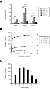
Figure 4. Influence of cytokines on sHLA-E production by tumor cells.
A/ sHLA-E detection in supernatants of three tumor cell lines: two melanoma cell lines (M88 and M102) and one colocarcinoma cell line (HT29), treated or not with IFN-α, IFN-γ or TNF-α (10 ng/ml, 48 h). Significant differences between the control and treatment values are indicated (*p<0.05, **p<0.01, ***p<0.001). B/ sHLA-E detection in culture supernatants of M102 treated with serial concentrations of IFN-α, IFN-γ and TNF-α for 48 h. C/ Time course of sHLA-E production in culture supernatant of M102 treated for up to 6 days with 10 ng/ml IFN-γ.

References

    1. Kim R, Emi M, Tanabe K. Cancer immunoediting from immune surveillance to immune escape. Immunology. 2007;121(1):1–14. - PMC - PubMed
    1. Marin R, Ruiz-Cabello F, Pedrinaci S, Mendez R, Jimenez P, et al. Analysis of HLA-E expression in human tumors. Immunogenetics. 2003;54(11):767–775. - PubMed
    1. Algarra I, Garcia-Lora A, Cabrera T, Ruiz-Cabello F, Garrido F. The selection of tumor variants with altered expression of classical and nonclassical MHC class I molecules: implications for tumor immune escape. Cancer Immunol Immunother. 2004;53(10):904–910. - PMC - PubMed
    1. Campoli M, Ferrone S. HLA antigen changes in malignant cells: epigenetic mechanisms and biologic significance. Oncogene. 2008;27(45):5869–5885. - PMC - PubMed
    1. Rouas-Freiss N, Moreau P, Ferrone S, Carosella ED. HLA-G proteins in cancer: do they provide tumor cells with an escape mechanism? Cancer Res. 2005;65(22):10139–10144. - PubMed

Publication types