. 2011 Jun 10:5:40.
doi: 10.3389/fnsys.2011.00040. eCollection 2011.

Nitric Oxide Synthase Inhibitor Improves De Novo and Long-Term l-DOPA-Induced Dyskinesia in Hemiparkinsonian Rats

Affiliations

Nitric Oxide Synthase Inhibitor Improves De Novo and Long-Term l-DOPA-Induced Dyskinesia in Hemiparkinsonian Rats

Fernando Eduardo Padovan-Neto et al. Front Syst Neurosci. .

Abstract

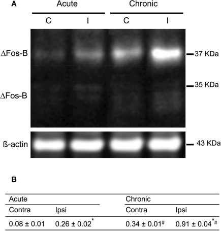
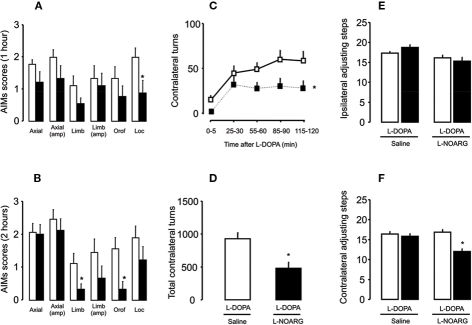
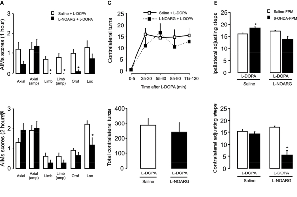
Inhibitors of neuronal and endothelial nitric oxide synthase decrease l-3,4-dihidroxifenilalanine (l-DOPA)-induced dyskinesias in rodents. The mechanism of nitric oxide inhibitor action is unknown. The aims of the present study were to investigate the decrease of l-DOPA-induced abnormal involuntary movements (AIMs) in 6-hydroxydopamine (6-OHDA)-lesioned rats by nitric oxide inhibitors following either acute or chronic treatment. The primary findings of this study were that NG-nitro-l-Arginine, an inhibitor of endothelial and neuronal nitric oxide synthase, attenuated AIMs induced by chronic and acute l-DOPA. In contrast, rotational behavior was attenuated only after chronic l-DOPA. The 6-OHDA lesion and the l-DOPA treatment induced a bilateral increase (1.5 times) in the neuronal nitric oxide synthase (nNOS) protein and nNOS mRNA in the striatum and in the frontal cortex. There was a parallel increase, bilaterally, of the FosB/ΔFosB, primarily in the ipsilateral striatum. The exception was in the contralateral striatum and the ipsilateral frontal cortex, where chronic l-DOPA treatment induced an increase of approximately 10 times the nNOS mRNA. Our results provided further evidence of an anti-dyskinetic effect of NOS inhibitor. The effect appeared under l-DOPA acute and chronic treatment. The l-DOPA treatment also revealed an over-expression of the neuronal NOS in the frontal cortex and striatum. Our results corroborated findings that l-DOPA-induced rotation differs between acute and chronic treatment. The effect of the NOS inhibitor conceivably relied on the l-DOPA structural modifications in the Parkinsonian brain. Taken together, these data provided a rationale for further evaluation of NOS inhibitors in the treatment of l-DOPA-induced dyskinesia.

Keywords: FosB/ΔFosB; Parkinson's disease; abnormal involuntary movements; l-DOPA-induced dyskinesia; nitric oxide; nitric oxide synthase; nitric oxide synthase inhibitors; striatum.


Figures

Figure 1
Figure 1
Representative microphotographs demonstrating TH-immunoreactive cell bodies in the substantia nigra of a rat with more than 95% cell loss. (A,B) The 6-OHDA-microinjection induced degeneration of dopamine neurons in the MFB injection side. Images were captured in sections at (A) −5.20 mm and (B) −6.04 mm, AP to Bregma. (C,D) Quantification of striatal TH + cells/fibers by optical density were performed at SNc (−5.20 mm from Bregma) and in the SNL and SNV (−6.04 mm from Bregma). Values are expressed as mean ± SEM (Saline-MFB, n = 6; 6-OHDA-MFB, n = 11). SNc, substantia nigra pars compacta; SNL, lateral substantia nigra; SNV, ventral substantia nigra.
Figure 2
Figure 2
Effects of 6-OHDA-lesion and l-DOPA treatment in the rodents stepping test. Rats received either saline (n = 17) or 6-OHDA (n = 11) microinjections on MFB. (A) For both groups, l-DOPA chronic treatment had no effect on the paw ipsilateral to the lesion. (B) 6-OHDA microinjection induced deficits in the paw contralateral to lesion (day 1, pre-test) that was reversed by l-DOPA administration (day 1, test). l-DOPA treatment also improved the akinesia in the contralateral paw until the end of the treatment (day 15). (C) Spearman correlation between contralateral adjusting steps as a function of indirect striatal dopamine depletion provided by apomorphine-induced rotational behavior. *p < 0.05 vs saline-FPM injected rats; #p < 0.05 vs pre-test (two way rANOVA). Values are expressed as mean ± SEM.
Figure 3
Figure 3
Quantitative real-time PCR analysis of the nNOS mRNA in the prefrontal cortex, striatum and hippocampus of control, 6-OHDA-lesioned and on established l-DOPA-induced AIMs. Treatment is described in the Section “Materials and Methods.” Bars represent transcript amount per region analyzed, expressed as the mean ± SEM of six to seven animals samples/group. Representative distribution patterns of nNOS mRNA expression on prefrontal cortex, striatum, and hippocampus are shown in (A), (B), and (C) respectively. p < 0.05 vs control; *p < 0.05 vs control and 6-OHDA-lesioned rats; #p < 0.05 vs contralateral side (rANOVA followed by the Sidak test, p < 0.05).
Figure 4
Figure 4
Western blot analysis of nNOS protein in the rat striatum: effect of lesion and l-DOPA chronic treatment in control, 6-OHDA-lesioned and on established l-DOPA-induced AIMs. Treatment is described in Methods. (A) Contralateral and, (B) ipsilateral striatum. Lines indicate specific bands at ∼155 kDa (nNOS) and ∼43 kDa of a loading control (β-actin). (C) There was an increment of nNOS protein expression in both ipsilateral and contrataleral striatum for 6-OHDA-lesioned and l-DOPA-treated-6-OHDA-lesioned rats. The values obtained from contralateral side were higher than the ipsilateral one. *p < 0.05 vs control; #p < 0.05 vs ipsilateral side (rANOVA followed by the Sidak test, p < 0.05).
Figure 5
Figure 5
Characterization of the expression of FosB/ΔFosB after acute and on established l-DOPA-induced AIMs. (A) Western blots of proteins from extracted striatum of acutely and chronically l-DOPA-treated hemiparkinsonian rats; left and right lanes represent proteins from ipsilateral (I) and contralateral (C) striatum, respectively. (B) Results of the quantification of l-DOPA-induced FosB/ΔFosB expression are expressed as the integrated density of each band of the proteins of interest was normalized to the integrated density of the control loading ß-actin (mean ± SEM; square pixel). *p < 0.05 compared with contralateral striatum for either acute or chronic treatment with l-DOPA (paired t-test). #p < 0.05 compared with the same side for acute or chronic treatment with l-DOPA (t-test).
Figure 6
Figure 6
Effects of l-NOARG pre-treatment on AIMs, rotational behavior and stepping test on established l-DOPA-induced AIMs. (A) l-NOARG pre-treatment significantly reduced locomotor AIM scores at 1 h after l-DOPA. (B) Limb and orofacial AIMs scores were significantly reduced by l-NOARG at 2 h after l-DOPA. *p < 0.05 (Wilcoxon). (C) l-NOARG pre-treatment reduced contralateral turns measured during 5 min at 0, 25, 55, 85, and 115 min after l-DOPA treatment. *p < 0.05 (one way rANOVA). (D) l-NOARG reduced total contralateral turns measured during 120 min after l-DOPA. p < 0.05 (paired t-test). After chronic l-DOPA treatment l-NOARG pre-treatment did not affect (E) ipsilateral adjusting steps and (F) slight reduced adjusting steps performed with contralateral paw. *p < 0.05 vs Saline-FPM (one way rANOVA). Values are expressed as mean ± SEM (6-OHDA-FPM, n = 8–9/group; Saline-FPM, n = 5–6/group).
Figure 7
Figure 7
Effects of l-NOARG pre-treatment on AIMs, rotational behavior and stepping test on acute l-DOPA-induced AIMs. (A) l-NOARG pre-treatment significantly reduced limb, limb amplitude and orofacial AIMs scores 1 h after l-DOPA application. (B) Locomotor AIM score was attenuated by l-NOARG 2 h after l-DOPA treatment. *p < 0.05 (Mann–Whitney). (C) l-NOARG pre-treatment had no effects on contralateral turns measured during 5 min at 0, 25, 55, 85, and 115 min after l-DOPA treatment p > 0.05 (two way rANOVA) neither on (D) total contralateral turns, assessed during 120 min after l-DOPA treatment. p > 0.05 (t-test) (E) l-DOPA treatment slightly increased the number of the adjusting steps of 6-OHDA-lesioned rats in the paw ipsilateral to the lesion. (F) l-DOPA treatment completely restored the akinesia in the contralateral-to lesion paw, of 6-OHDA-lesioned rats but l-NOARG pre-treatment impaired l-DOPA effect. *p < 0.05 vs Saline-FPM injected (two way ANOVA). Values are expressed as mean ± SEM (6-OHDA-FPM, n = 10–11/group; Saline-FPM, n = 5/group).

References

    1. Abreu-González P., González-Hernández T., Afonso-Oramas D., Cruz-Muros I., Barroso-Chinea P., González M. C. (2006). Tetrahydrobiopterin stimulates l-DOPA release from striatal tissue. Eur. J. Pharmacol. 541, 33–37 - PubMed
    1. Andersson M., Hilbertson A., Cenci M. A. (1999). Striatal fosB expression is causally linked with l-DOPA-induced abnormal involuntary movements and the associated upregulation of striatal prodynorphin mRNA in a rat model of Parkinson's disease. Neurobiol. Dis. 6, 461–474 - PubMed
    1. Aquilano K., Baldelli S., Rotilio G., Ciriolo MR. (2008). Role of nitric oxide synthases in Parkinson's disease: a review on the antioxidant and anti-inflammatory activity of polyphenols. Neurochem. Res. 33, 2416–2426 - PubMed
    1. Barone P. (2010). Neurotransmission in Parkinson's disease: beyond dopamine. Eur. J. Neurol. 17, 364–376 - PubMed
    1. Barthwal M. K., Srivastava N., Dikshit M. (2001). Role of nitric oxide in a progressive neurodegeneration model of Parkinson's disease in the rat. Redox Rep. 6, 297–30210.1179/135100001101536436 - DOI - PubMed