. 2011 Jun 6:5:4.
doi: 10.3389/fncel.2011.00004. eCollection 2011.

Understanding the molecular diversity of GABAergic synapses

Affiliations

Understanding the molecular diversity of GABAergic synapses

Marco Sassoè-Pognetto et al. Front Cell Neurosci. .

Abstract

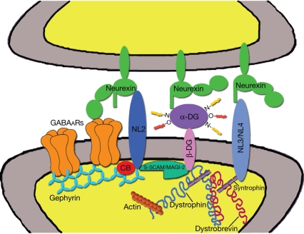
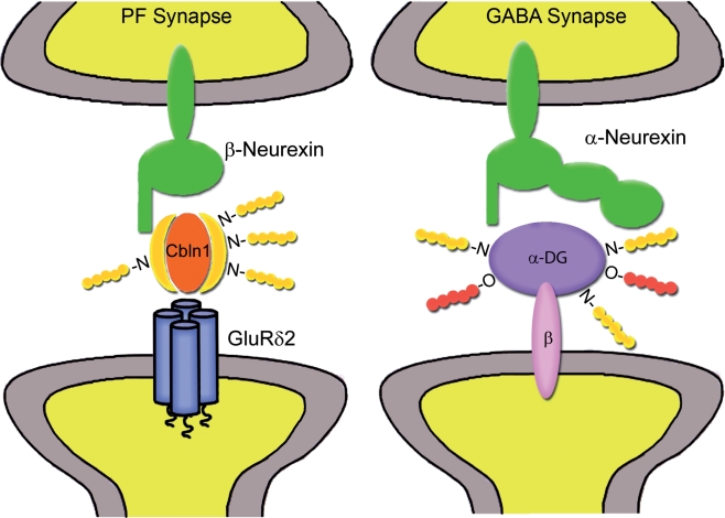
GABAergic synapses exhibit a high degree of subcellular and molecular specialization, which contrasts with their apparent simplicity in ultrastructural appearance. Indeed, when observed in the electron microscope, GABAergic synapses fit in the symmetric, or Gray's type II category, being characterized by a relatively simple postsynaptic specialization. The inhibitory postsynaptic density cannot be readily isolated, and progress in understanding its molecular composition has lagged behind that of excitatory synapses. However, recent studies have brought significant progress in the identification of new synaptic proteins, revealing an unexpected complexity in the molecular machinery that regulates GABAergic synaptogenesis. In this article, we provide an overview of the molecular diversity of GABAergic synapses, and we consider how synapse specificity may be encoded by selective trans-synaptic interactions between pre- and postsynaptic adhesion molecules and secreted factors that reside in the synaptic cleft. We also discuss the importance of developing cataloguing tools that could be used to decipher the molecular diversity of synapses and to predict alterations of inhibitory transmission in the course of neurological diseases.

Keywords: GABAA receptor; GABAergic synapse; dystroglycan; gephyrin; neurexin; neuroligin; synapse specificity.


Figures

Figure 1
Figure 1
Postsynaptic scaffolds and adhesion molecules of GABAergic synapses. The diagram is based on reported molecular interactions (see text), some of which remain to be confirmed in vivo. Gephyrin, S-SCAM/MAGI-2, and dystrophin are shown in the same postsynaptic specialization, although dystrophin is present in only a subset of GABAergic synapses and the in vivo distribution of S-SCAM/MAGI-2 has not been characterized. Gephyrin trimers are believed to aggregate into a submembranous lattice that provides stability to postsynaptic GABAARs. Gephyrin also binds collybistin (CB) and neuroligin 2 (NL2), that has been proposed to function as a specific activator of collybistin. Cytoskeletal proteins associated with gephyrin, such as Mena/VASP and microtubules, are not shown. Neuroligin 2 bridges the synaptic cleft and binds to neurexins on the presynaptic terminal. Reported interactions between neurexins and GABAARs are also indicated. Neurexins may also interact with α-dystroglycan (α-DG), thus establishing a link with the dystrophin–glycoprotein complex (DGC). One component of the DGC, syntrophin, has been reported to bind neuroligin 3 and neuroligin 4. Finally, there is evidence that S-SCAM/MAGI-2 may establish a link between neuroligin 2 and the DGC by interacting with the intracellular domain of β-dystroglycan (β-DG).
Figure 2
Figure 2
Proposed trans-synaptic interactions mediated by synaptic cleft glycoproteins. At parallel fiber-Purkinje cell synapses (left), Cbln1 forms a ternary complex with neurexin variants containing the S4 insert and postsynaptic GluRδ2 receptors (modified from Uemura et al., 2010). At some GABAergic synapses (right), α-dystroglycan (α-DG) may establish a link between α-neurexins and the postsynaptic dystrophin–glycoprotein complex through the transmembrane β-dystroglycan isoform (β-DG). Glycan side chains of Cbln1 and α-DG may also mediate multiple interactions with extracellular matrix molecules.

References

    1. Alldred M. J., Mulder-Rosi J., Lingenfelter S. E., Chen G., Luscher B. (2005). Distinct γ2 subunit domains mediate clustering and synaptic function of postsynaptic GABAA receptors and gephyrin. J. Neurosci. 25, 594–60310.1523/JNEUROSCI.4011-04.2005 - DOI - PMC - PubMed
    1. Anderson J. L., Head S. I., Morley J. W. (2003). Altered inhibitory input to Purkinje cells of dystrophin-deficient mice. Brain Res. 982, 280–28310.1016/S0006-8993(03)03018-X - DOI - PubMed
    1. Ango F., Di Cristo G., Higashiyama H., Bennett V., Wu P., Huang Z. J. (2004). Ankyrin-based subcellular gradient of neurofascin, an immunoglobulin family protein, directs GABAergic innervation at Purkinje axon initial segment. Cell 119, 257–27210.1016/j.cell.2004.10.004 - DOI - PubMed
    1. Ango F., Wu C., Van der Want J. J., Wu P., Schachner M., Huang Z. J. (2008). Bergmann glia and the recognition molecule CHL1 organize GABAergic axons and direct innervation of Purkinje cell dendrites. PLoS Biol. 6, e103.10.1371/journal.pbio.0060103 - DOI - PMC - PubMed
    1. Arikkath J., Reichardt L. F. (2008). Cadherins and catenins at synapses: roles in synaptogenesis and synaptic plasticity. Trends Neurosci. 31, 487–49410.1016/j.tins.2008.07.001 - DOI - PMC - PubMed