. 2011 Jun 8:5:32.
doi: 10.3389/fnsys.2011.00032. eCollection 2011.

An Improved BAC Transgenic Fluorescent Reporter Line for Sensitive and Specific Identification of Striatonigral Medium Spiny Neurons

Affiliations

An Improved BAC Transgenic Fluorescent Reporter Line for Sensitive and Specific Identification of Striatonigral Medium Spiny Neurons

Kristen K Ade et al. Front Syst Neurosci. .

Abstract

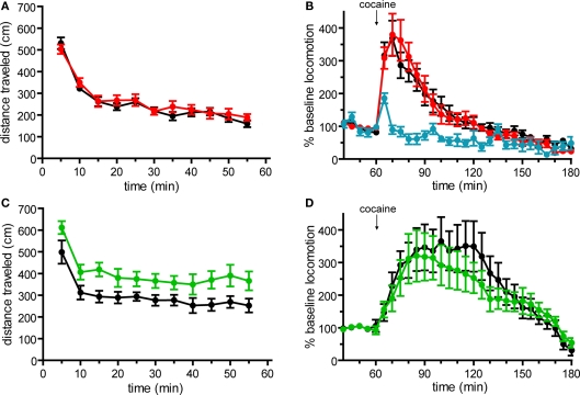
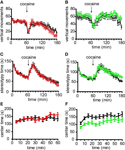
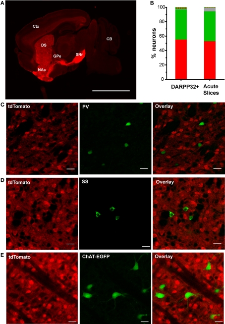
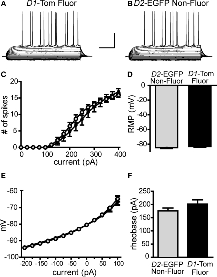
The development of BAC transgenic mice expressing promoter-specific fluorescent reporter proteins has been a great asset for neuroscience by enabling detection of neuronal subsets in live tissue. For the study of basal ganglia physiology, reporters driven by type 1 and 2 dopamine receptors have been particularly useful for distinguishing the two classes of striatal projection neurons - striatonigral and striatopallidal. However, emerging evidence suggests that some of the transgenic reporter lines may have suboptimal features. The ideal transgenic reporter line should (1) express a reporter with high sensitivity and specificity for detecting the cellular subset of interest and that does not otherwise alter the biology of the cells in which it is expressed, and (2) involve a genetic manipulation that does not cause any additional genetic effects other than expression of the reporter. Here we introduce a new BAC transgenic reporter line, Drd1a-tdTomato line 6, with features that approximate these ideals, offering substantial benefits over existing lines. In this study, we investigate the integrity of dopamine-sensitive behaviors and test the sensitivity and specificity of tdTomato fluorescence for identifying striatonigral projection neurons in mice. Behaviorally, hemizygous Drd1a-tdTomato line 6 mice are similar to littermate controls; while hemizygous Drd2-EGFP mice are not. In characterizing the sensitivity and specificity of line 6 mice, we find that both are high. The results of this characterization indicate that line 6 Drd1a-tdTomato+/- mice offer a useful alternative approach to identify both striatonigral and striatopallidal neurons in a single transgenic line with a high degree of accuracy.

Keywords: BAC; basal ganglia; direct pathway; medium spiny neuron; striatonigral; striatum; tdTomato; transgenic.


Figures

Figure 1
Figure 1
Basal locomotor activity and acute locomotor response to cocaine in Drd1a-tdTomato line 6 and Drd2-EGFP BAC transgenic mice on C57Bl/6 background. (A) Basal horizontal locomotion of Drd1a-tdTomato+/− line 6 mice (red, n = 14) is similar to transgene-negative (Drd1a-tdTomato−/−) littermates (black, n = 15; p = 0.515). (B) Acute cocaine administration (20 mg/kg i.p.) similarly increases horizontal locomotion in Drd1a-tdTomato+/− mice (red, n = 14) and Drd1a-tdTomato−/− littermates (black, n = 15; p = 0.945). Blue trace indicates response to vehicle control injection (n = 8 mice, 4 each of Drd1a-tdTomato−/− and Drd1a-tdTomato+/− mice. Responses were indistinguishable, so data were aggregated.) (C) Time course plots of basal horizontal locomotion demonstrate that Drd2-EGFP+/− mice (green, n = 17) are hyperactive compared to transgene-negative (Drd2-EGFP−/−) littermates (black, n = 15; p = 0.027). (D) Acute cocaine administration (20 mg/kg i.p.) induces a similar percent increase in locomotion in Drd2-EGFP+/− mice (green, n = 17) and Drd2-EGFP−/− littermates (black, n = 15; p = 0.616).
Figure 2
Figure 2
Non-locomotor behaviors in line 6 Drd1a-tdTomato/C57Bl/6 and Drd2-EGFP/C57Bl/6 transgenic mice. Time course plots of the number of vertical movements basally and following acute cocaine injection (20 mg/kg i.p.) demonstrate that transgene-positive mice are similar to transgene-negative littermate control mice in (A) Drd1a-tdTomato line 6 mice (Drd1a-tdTomato+/−, red, n = 14; Drd1a-tdTomato−/−, black, n = 15; p = 0.778) and (B) Drd2-EGFP mice (Drd2-EGFP+/−, green, n = 17; Drd2-EGFP−/−, black, n = 15; p = 0.256). No differences in stereotypic movements basally and following acute cocaine injection were observed between transgene-positive and transgene-negative littermate control mice in (C) Drd1a-tdTomato line 6 mice (Drd1a-tdTomato+/−, red, n = 14; Drd1a-tdTomato−/−, black, n = 15; p = 0.615) and (D) Drd2-EGFP mice (Drd2-EGFP+/−, green, n = 17; Drd2-EGFP−/−, black, n = 15; p = 0.411). (E) Drd1a–tdTomato line 6 mice and transgene-negative littermate control mice spend a similar amount of time in center region of open field test chamber (Drd1a-tdTomato+/−, red, n = 14; Drd1a-tdTomato−/−, black, n = 15; p = 0.647). (F) Drd2-EGFP+/− (green, n = 17) spend significantly less time in the center region of the open field chamber compared to Drd2-EGFP−/− littermates (black, n = 15; p = 0.013).
Figure 3
Figure 3
TdTomato fluorescence is sensitive and specific for striatonigral MSNs in the dorsal striatum. (A) A representative low-magnification sagittal view of tdTomato fluorescence of a brain slice from a hemizygous line 6 Drd1a-tdTomato mouse demonstrating strong signal within striatal efferent axons terminating in substantia nigra pars reticulata (SNr) and negligible signal at the globus pallidus external (GPe). Abbreviations: cortex (Ctx), cerebellum (CB), dorsal striatum (DS) and nucleus accumbens (NAc). Scale bar represents 500 μm. (B) Quantification of fluorescent signal detected in dorsal striatal cells from line 6 Drd1a-tdTomato+/−/Drd2-EGFP+/− mice. DARPP32+ cells were identified in immunohistochemically stained 50 μm thick brain sections (n = 605 tdTomato only (red), 457 EGFP only (green), 25 dually labeled (red and green striped), and 8 non-fluorescent (gray) of 1095 total DARPP-32 immunopositive cells). Cells in acute 300 μm thick slices were identified by phase-contrast illumination. Fluorescent signals were detected using epifluorescence illumination (n = 293 tdTomato only (red), 230 EGFP only (green), 5 dually labeled (red and green striped), and 26 non-fluorescent (gray) of 549 total medium size cells identified by phase contrast illumination). (C) Representative confocal microscopy images demonstrating that cells immunopositive for the fast-spiking GABAergic interneuron marker, parvalbumin (PV), do not express tdTomato (0/52 PV-immunopositive cells). (D) Representative confocal microscopy images demonstrating cells immunopositive for the low-threshold spiking GABAergic interneuron marker, somatostatin (SS), do not express tdTomato (0/45 SS-immunopositive cells). (E) Representative confocal microscopy images demonstrating no overlap between tdTomato and EGFP fluorescence in line 6 Drd1a-tdTomato+/−/ChAT-EGFP+/− mice (0/56 EGFP-positive cells). For panels (C–E), tdTomato fluorescence (left), interneuron antigen or transgenic reporter fluorescence (middle) and overlay of both signals (right). Scale bars represent 25 μm.
Figure 4
Figure 4
Expression of tdTomato reporter does not affect intrinsic membrane properties in striatonigral pathway MSNs. Whole-cell, current clamp recordings of striatonigral MSNs identified by tdTomato fluorescence in line 6 Drd1a-tdTomato+/−/Drd2-EGFP+/− mice (D1-Tom Fluor) or by lack of EGFP fluorescence in Drd1a-tdTomato−/−/Drd2-EGFP+/− littermates (D2-EGFP Non-Fluor) reveals no significant differences between the two groups. Representative responses from current clamp recordings in response to a series of current injections (25 pA increments) are shown for (A) a D1-Tom Fluor direct pathway MSN and (B) a D2-EGFP Non-Fluor direct pathway MSN. Summary graphs show D1-Tom Fluor MSNs (shown in black/closed circles, n = 12) and D2-EGFP Non-Fluor MSNs (shown in gray/open circles, n = 12) do not differ in (C) number of action potential spikes fired in response to 500-ms duration current injection steps, (D) rheobase, (E) current–voltage relationship/ input resistance or (F) resting membrane potential (RMP).
Figure 5
Figure 5
Survey of tdTomato expression in sagittal brain slices prepared from line 6 Drd1a-tdTomato+/− mice. Representative confocal images showing tdTomato expression at (A) the border between cortex and striatum (corpus callosum = CC), (B) the nucleus accumbens (NAc) and (C) the cerebellum (Purkinje cell = PC). (D) Representative epifluorescent image showing tdTomato expression in the hippocampus (dentate gyrus = DG). The contrast has been enhanced for each image to highlight tdTomato expression patterns within the subregion. Scale bars for all panels represent 100 μm.

References

    1. Ade K. K., Janssen M. J., Ortinski P. I., Vicini S. (2008). Differential tonic GABA conductances in striatal medium spiny neurons. J. Neurosci. 28, 1185–119710.1523/JNEUROSCI.3908-07.2008 - DOI - PMC - PubMed
    1. Anderson K. D., Reiner A. (1991). Immunohistochemical localization of DARPP-32 in striatal projection neurons and striatal interneurons: implications for the localization of D1-like dopamine receptors on different types of striatal neurons. Brain Res. 568, 235–24310.1016/0006-8993(91)91403-N - DOI - PubMed
    1. André V. M., Cepeda C., Fisher Y. E., Huynh M., Bardakjian N., Singh S., Yang X. W., Levine M. S. (2011). Differential electrophysiological changes in striatal output neurons in Huntington's disease. J. Neurosci. 31, 1170–118210.1523/JNEUROSCI.3539-10.2011 - DOI - PMC - PubMed
    1. Bateup H. S., Santini E., Shen W., Birnbaum S., Valjent E., Surmeier D. J., Fisone G., Nestler E. J., Greengard P. (2010). Distinct subclasses of medium spiny neurons differentially regulate striatal motor behaviors. Proc. Natl. Acad. Sci. U.S.A. 107, 14845–14850 - PMC - PubMed
    1. Bertran-Gonzalez J., Bosch C., Maroteaux M., Matamales M., Herve D., Valjent E., Girault J. A. (2008). Opposing patterns of signaling activation in dopamine D1 and D2 receptor-expressing striatal neurons in response to cocaine and haloperidol. J. Neurosci. 28, 5671–568510.1523/JNEUROSCI.1039-08.2008 - DOI - PMC - PubMed