. 2011 Jun 8:5:27.
doi: 10.3389/fnbeh.2011.00027. eCollection 2011.

High-precision, three-dimensional tracking of mouse whisker movements with optical motion capture technology

Affiliations

High-precision, three-dimensional tracking of mouse whisker movements with optical motion capture technology

Snigdha Roy et al. Front Behav Neurosci. .

Abstract

The mystacial vibrissae or whiskers in rodents are sensitive tactile hairs emerging from both sides of the face. Rats and mice actively move these whiskers during exploration. The neuronal mechanisms controlling whisker movements and the sensory representation of whisker tactile information are widely studied as a model for sensorimotor processing in mammals. Studies of the natural whisker movement patterns during exploration and tactile examination are still in their early stages. Tracking the movements of whiskers is technically challenging as they move relatively fast and are very thin, particularly in mice. Existing systems detect light-beam interruptions by the whiskers or use high-speed video to track whisker movements in one or two-dimensions. Here we describe a method for tracking the movements of mouse whiskers in three-dimensions (3D) using optical motion capture technology (OMCT). OMCT tracks the movements of small retro-reflective markers attached to whiskers of a head-fixed mouse with a spatial resolution of <0.5 mm in all 3D and a temporal resolution of 5 ms (200 fps). The system stores the 3D coordinates of the marker's trajectories onto hard disk allowing a detailed analysis of movement trajectories bilateral coordination. The described method currently uses the minimum of two tracking cameras, which requires head-fixation for reliable tracking.

Keywords: 3D tracking; mouse; movement tracking; mystacial vibrissae; whisker movement.


Figures

Figure 1
Figure 1
Arrangement of tracking system, placement of whisker markers and virtual representation. (A) Photograph showing two high-speed cameras (on the left) with infrared lights are placed 0.8–1.2 m from the mouse (bottom right), with cameral sightlines converging on the mouse at an approximate 24° angle. The mouse's head is immobilized with a head post. (B) Photographs showing close-up front and side-views of retro-reflective marker tape affixed to the left (green arrows) and right (purple arrow) C4 whisker of a C57BL/6J mouse. Acrylic cement used to mount the head post is partially visible on to of the mouse's head. (C) Screenshot from the Cortex Software during calibration showing the positions of the two-cameras (green boxes) relative to each other, outlines of their fields of view and the volume viewed by both cameras for 3D tracking. (D) Screenshot from the Cortex Software during the calibration process showing the digital representation of three retro-reflective markers, one static and two attached to the left and right C4 whiskers.
Figure 2
Figure 2
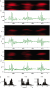
Spectral analysis of whisker movements. (A) Spectrograms (black and red heat-maps) and trajectory components for whisker movements along each axis (green traces) are shown. Spectrograms (Hanning, block size 256) for whisker movements along all three axes show high power (bright red colors) within a stable frequency band centered around 15 Hz during whisker movements with variable amplitude. (B) Power spectral density histograms (Hanning, FFT size 256) calculated for the length of the trajectory components in (A) show prominent peaks around 15 Hz.
Figure 3
Figure 3
Different viewing angles for three-dimensional (3D) reconstructions of C4 whisker movements. (A) Frontal view of the marker positions during 100 s (20000 frames) of whisker movements. Markers were attached to the left and right C4 whiskers. Black dots in this and all other plots represent 3D marker positions. Red dots are projections of the 3D positions onto each of the three planes. (B) Side-view of (A) rotated 35° to the right. (C) Side-view of (A) rotated 90° to the right. (D) Top down view (along the z-axis) of (C).
Figure 4
Figure 4
Example raw traces of the X, Y, and Z components of bilaterally symmetrical and non-symmetrical whisker movements. (A) Traces showing from top to bottom the X, Y, and Z (top to bottom) components of three-dimensional movements of the left (green traces) and right (gray traces) C4 whiskers. During the 2-s (400 frames) of recordings shown the whiskers moved in a bilaterally symmetrical way. (B) Table with correlation coefficients from a correlation analysis of X, Y, and Z components of bilateral whisker movements shows positive correlations between corresponding components (red framed values along diagonal). (C) Traces as in (A) recorded in the same session but during a 2-s period of non-symmetrical whisker movements. (D) Correlation analysis of X, Y, and Z components of bilateral whisker movements shows negative correlations between corresponding components (red framed values along diagonal).

References

    1. Abdel-Aziz Y. I., Karara H. M. (1971). “Direct linear transformation from comparator coordinates into object-space coordinates in close-range photogrammetry,” in Proceedings of the ASP/UI Symposium on Close-Range Photogrammetry (Falls Church, VA: American Society of Photogrammetry), 1–18
    1. Berg R. W., Kleinfeld D. (2003). Rhythmic whisking by rat: retraction as well as protraction of the vibrissae is under active muscular control. J. Neurophysiol. 89, 104–11710.1152/jn.00600.2002 - DOI - PubMed
    1. Bermejo R., Houben D., Zeigler H. P. (1998). Optoelectronic monitoring of individual whisker movements in rats. J. Neurosci. Methods 83, 89–96 - PubMed
    1. Blake R. L., Ferguson H. J. (1993). The motion analysis system for dynamic gait analysis. Clin. Podiatr. Med. Surg. 10, 501–527 - PubMed
    1. Bryant J. L., Roy S., Heck D. H. (2009). A technique for stereotaxic recordings of neuronal activity in awake, head-restrained mice. J. Neurosci. Methods 178, 75. - PMC - PubMed