

Choroidal metastasis from carcinoma of breast detected on F18-FDG PET CT scan: A case report and review of literature
- PMID: 21713225
- PMCID: PMC3109823
- DOI: 10.4103/0972-3919.78252
Choroidal metastasis from carcinoma of breast detected on F18-FDG PET CT scan: A case report and review of literature
Abstract
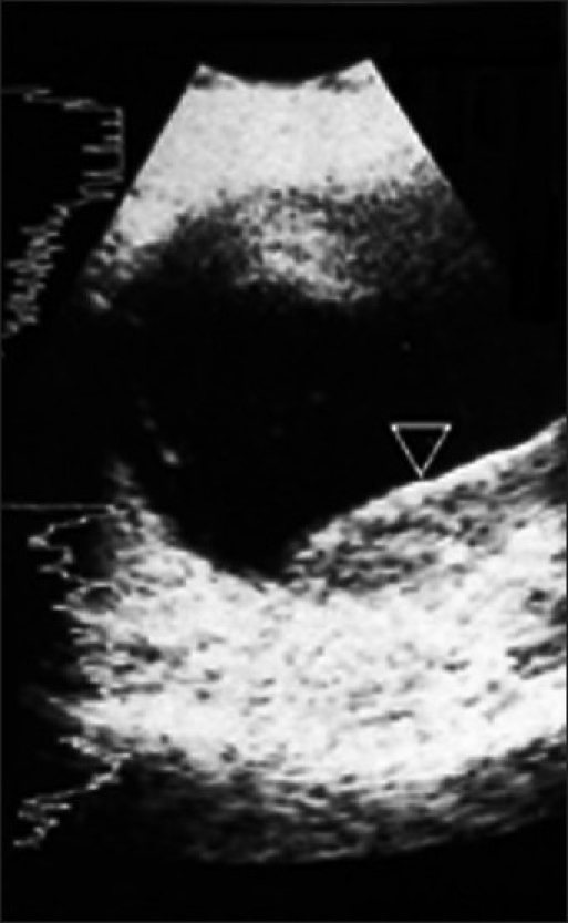
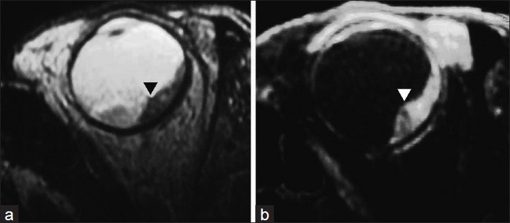
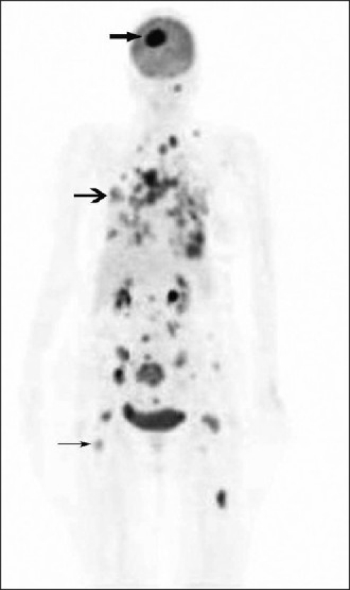
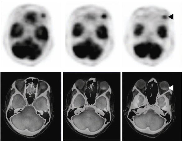
Intraocular choroidal metastasis is a very rare cause of blindness. Choroidal hemangioma and melanoma are other causes that may mimic the condition. Carcinoma of breast is the most common primary malignancy that accounts for choroidal metastasis in females and carcinoma of lung is the most common cause in males. Other primary neoplasms which can uncommonly metastasize to the choroid are testis, gastrointestinal tract, kidney, thyroid, pancreas, and prostate. Metastatic neoplasm to the eye outnumbers the primary tumors such as retinoblastomas and malignant melanoma. Sonography is usually the initial investigation after fundus examination to look for the architecture of the lesion. However, it lacks in specificity. We present a case of carcinoma of breast that had visual disturbances and wholebody F18-fluorodeoxyglucose, positron emission tomography-computerized tomography (FDG PET CT) revealed a choroidal lesion in addition to cerebral, pulmonary, and skeletal metastases. Choroidal metastasis from carcinoma of lung has been reported previously on FDG PET. To the best of our knowledge, this is the first case report of carcinoma of breast demonstrating choroid metastasis on F18-FDG PET CT scan.
Keywords: Distant metastasis; paranasal sinuses; thyroid neoplasms.
Conflict of interest statement
Figures
References
-
- Morton KA, Clark PB. Diagnostic imaging- Nuclear medicine. 1st ed. Utah, USA: Amirsys inc. Salt Lake; 2007. pp. 11–35.
-
- Mewis L, Young SE. Breast carcinoma metastatic to the choroid: Analysis of 67 patients. Ophthalmology. 1982;89:147–51. - PubMed
-
- Shields CL, Shields JA, Gross NE, Schwartz GP, Lally SE. Survey of 520 eyes with uveal metastases. Ophthalmology. 1997;104:1265–6. - PubMed
-
- Demirci H, Shields CL, Chao A, Shields JA. Uveal metastasis from breast cancer in 264 patients. Am J Ophthalmol. 2003;136:264–71. - PubMed
-
- Shields JA, Shields CL, Kiratli H, de Potter P. Metastatic tumors to the iris in 40 patients. Am J Ophthalmol. 1995;119:422–30. - PubMed
