Review
. 2011 Nov;71(11):901-23.
doi: 10.1002/dneu.20931.

Regulation of axonal outgrowth and pathfinding by integrin-ECM interactions

Affiliations
Review

Regulation of axonal outgrowth and pathfinding by integrin-ECM interactions

Jonathan P Myers et al. Dev Neurobiol. 2011 Nov.

Abstract

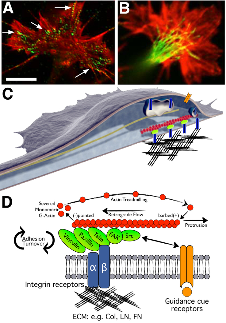
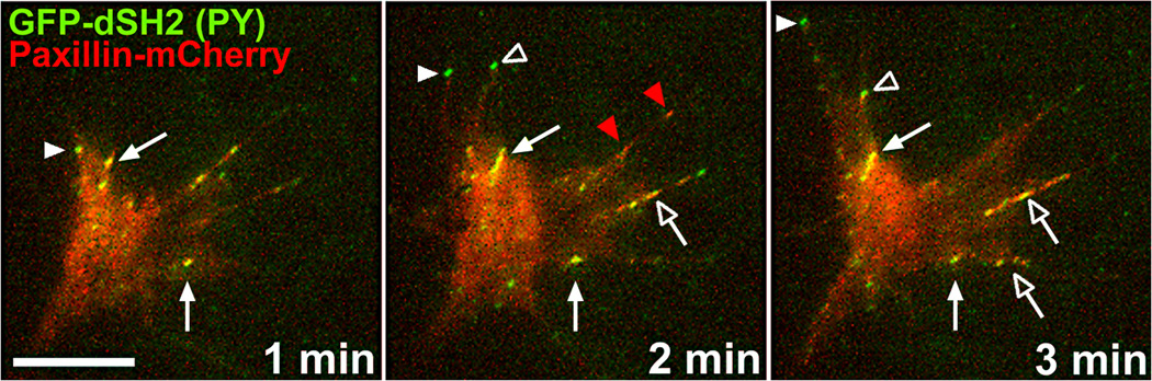
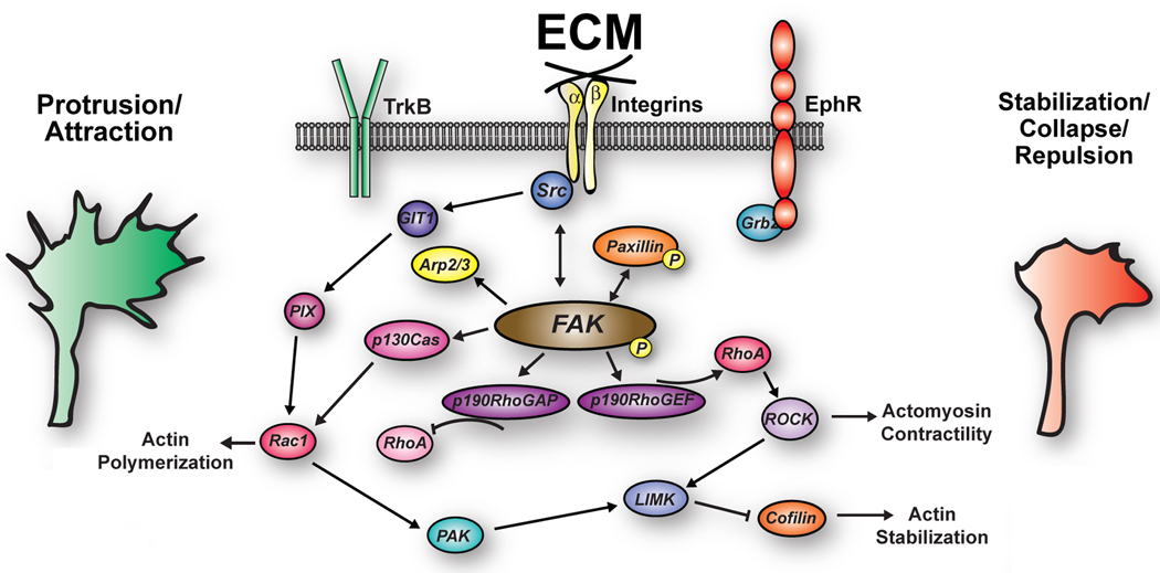
Developing neurons use a combination of guidance cues to assemble a functional neural network. A variety of proteins immobilized within the extracellular matrix (ECM) provide specific binding sites for integrin receptors on neurons. Integrin receptors on growth cones associate with a number of cytosolic adaptor and signaling proteins that regulate cytoskeletal dynamics and cell adhesion. Recent evidence suggests that soluble growth factors and classic axon guidance cues may direct axon pathfinding by controlling integrin-based adhesion. Moreover, because classic axon guidance cues themselves are immobilized within the ECM and integrins modulate cellular responses to many axon guidance cues, interactions between activated receptors modulate cell signals and adhesion. Ultimately, growth cones control axon outgrowth and pathfinding behaviors by integrating distinct biochemical signals to promote the proper assembly of the nervous system. In this review, we discuss our current understanding how ECM proteins and their associated integrin receptors control neural network formation.


Figures

Figure 1
Figure 1. Growth cones assemble macromolecular adhesion complexes (point contacts) that link ECM proteins to actin filaments
A. A Xenopus spinal neuron growth cone immuno-labelled for phospho-Tyr118-paxillin (green) and F-actin (red). Note multiple point contact adhesions along actin filaments (arrows). B. A Xenopus spinal neuron growth cone immuno-labelled for β-tubulin (green) and F-actin (red). Note that microtubules may regulate the trafficking of vesicles containing integrin and guidance cue receptors. Scale, 5 µm. C. Schematic representation of a growth cone (adapted from (Kamiguchi and Lemmon, 2000)) on the ECM with several integrin receptors (blue) linked to actin filaments through adhesion complexes (green). Integrin receptor trafficking within recycling endosomes (blue vesicles) along microtubules (dark green) may regulate axon guidance (see text for details) A guidance cue/growth factor receptor is illustrated on the apical surface (orange). D. Schematic representation of key molecular components of growth cone point contact adhesions. Integrin αβ heterodimeric receptors (dark blue lines) bind to proteins within the ECM, such as Col, LN and FN. Integrin activation leads to the assembly of multiple scaffolding proteins, such as talin, paxillin and vinculin to the cytoplasmic tail of integrins. In addition, FAK and Src are activated by clustering of integrin receptors, and they modulate the composition of adhesions through phosphorylation of key residues that allow for binding of many additional proteins (not shown). Several scaffolding proteins bind directly to actin filaments (red), which is believed to restrain retrograde flow and allow the force of actin polymerization to generate membrane protrusion. Guidance cue receptors (orange) can also regulate adhesion-associated proteins through binding and activation of FAK and Src. Cross-talk through FAK/Src signaling modulates adhesion assembly and turnover, as well as regulation of the actin cytoskeleton.
Figure 2
Figure 2. Two-channel total internal reflection fluorescence (TIRF) microscopy of phosphotyrosine (PY) and paxillin in a living growth cone
This neuron is co-expressing GFP-dSH2 (green), which brightly labels regions containing concentrated tyrosine-phosphorylated proteins and paxillin-mCherry (red), which labels integrin-dependent adhesion sites. Note that only PY is concentrated at the tips of elongating filopodia (solid white arrowheads mark one extending filopodium and open white arrowheads mark a filopodium that extends then retracts), which appear green due to the absence of paxillin. On the other hand, stable adhesions that contain both paxillin and PY appear yellow throughout this time period (solid arrows). Stable adhesion likely contain many other signaling and adaptor proteins, such as FAK and α-actinin. New adhesions often form from retracting PY-positive filopodia that stabilize and cluster with paxillin (at open arrowhead at 3 min) or by apparent simultaneous clustering of paxillin and PY-proteins within nascent protrusions (open arrows). However, in some instances, paxillin appears to cluster at adhesion sites independent of PY-containing proteins (red arrowheads). Scale, 10 µm.
Figure 3
Figure 3. Cooperation between integrin-dependent signaling pathways and modulatory receptors regulate growth cone motility by balancing Rho GTPase signaling
Integrins signal through a Src-FAK complex to promote membrane protrusion by elevating Rac1 and inhibiting RhoA. However, shifting this balance to more RhoA and less Rac1 activity leads to less actin polymerization and more actomyosin contractility. Modest RhoA signaling will stabilize adhesions to promote growth cone stalling or turning. Strong RhoA signaling will cause growth cone collapse and axon retraction. Shifting the balance of Rho GTPase signaling may occur through growth-promoting and inhibiting extracellular ligands that directly signal through Src-FAK. Alternatively, modulation of Src-FAK signals may occur as a result of re-association of FAK-associated protein complexes with modulating receptors.

References

    1. Ackley BD, Crew JR, Elamaa H, Pihlajaniemi T, Kuo CJ, Kramer JM. The NC1/endostatin domain of Caenorhabditis elegans type XVIII collagen affects cell migration and axon guidance. J Cell Biol. 2001;152:1219–1232. - PMC - PubMed
    1. Adams DN, Kao EY, Hypolite CL, Distefano MD, Hu WS, Letourneau PC. Growth cones turn and migrate up an immobilized gradient of the laminin IKVAV peptide. J Neurobiol. 2005;62:134–147. - PubMed
    1. Andrews MR, Czvitkovich S, Dassie E, Vogelaar CF, Faissner A, Blits B, Gage FH, ffrench-Constant C, Fawcett JW. Alpha9 integrin promotes neurite outgrowth on tenascin-C and enhances sensory axon regeneration. J Neurosci. 2009;29:5546–5557. - PMC - PubMed
    1. Anton ES, Kreidberg JA, Rakic P. Distinct functions of alpha3 and alpha(v) integrin receptors in neuronal migration and laminar organization of the cerebral cortex. Neuron. 1999;22:277–289. - PubMed
    1. Aumailley M, Bruckner-Tuderman L, Carter WG, Deutzmann R, Edgar D, Ekblom P, Engel J, Engvall E, Hohenester E, Jones JC, Kleinman HK, Marinkovich MP, Martin GR, Mayer U, Meneguzzi G, Miner JH, Miyazaki K, Patarroyo M, Paulsson M, Quaranta V, Sanes JR, Sasaki T, Sekiguchi K, Sorokin LM, Talts JF, Tryggvason K, Uitto J, Virtanen I, von der Mark K, Wewer UM, Yamada Y, Yurchenco PD. A simplified laminin nomenclature. Matrix Biol. 2005;24:326–332. - PubMed

Publication types