. 2012 Mar;22(2):150-8.
doi: 10.1111/j.1750-3639.2011.00514.x. Epub 2011 Sep 15.

Acute murine H5N1 influenza A encephalitis

Affiliations

Acute murine H5N1 influenza A encephalitis

Stephanie J Bissel et al. Brain Pathol. 2012 Mar.

Abstract

Avian influenza A virus H5N1 has the proven capacity to infect humans through cross-species transmission, but to date, efficient human-to-human transmission is limited. In natural avian hosts, animal models and sporadic human outbreaks, H5N1 infection has been associated with neurological disease. We infected BALB/c mice intranasally with H5N1 influenza A/Viet Nam/1203/2004 to study the immune response during acute encephalitis. Using immunohistochemistry and in situ hybridization, we compared the time course of viral infection with activation of immunity. By 5 days postinfection (DPI), mice had lost substantial body weight and required sacrifice by 7 DPI. H5N1 influenza was detected in the lung as early as 1 DPI, whereas infected neurons were not observed until 4 DPI. H5N1 infection of BALB/c mice developed into severe acute panencephalitis. Infected neurons lacked evidence of a perineuronal net and exhibited signs of apoptosis. Whereas lung influenza infection was associated with an early type I interferon (IFN) response followed by a reduction in viral burden concordant with appearance of IFN-γ, the central nervous system environment exhibited a blunted type I IFN response.


Figures

Figure 1
Figure 1
Weight loss and viral replication of influenza VN/04 in BALB/c mice following intranasal challenge. A. Weight loss was assessed daily following viral challenge. Mice that lost substantial amounts of their original body weight were euthanized. Viral burdens in the lungs (B) and the brain (C) were determined by plaque assays using Madin Darby canine kidney (MDCK) cells (n = 5/time point). Dashed line indicates limit of detection. Abbreviation: PFU = plaque‐forming unit.
Figure 2
Figure 2
Influenza in situ hybridization (ISH) shows infected cells in lung at 1 day postinfection (DPI) and in brain at 4 DPI. A. Representative ISH time course for influenza A MP is depicted (1–7 DPI) for lung (left column) and brain (right column). B. Severity of influenza ISH foci was accessed daily in sagittal sections of brains (n = 5/time point). Scoring: 0 = no definitive signal, 1 = occasional focus, 2 = focus in most fields, 3 = more than one focus per field.
Figure 3
Figure 3
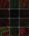
Influenza staining is observed in neurons lacking perineuronal nets. A–C. Double‐label immunofluorescent stains of sagittal sections from 6 or 7 days postinfection show that neurons, not microglia or astrocytes, were the predominant infected cell. Influenza A staining is shown in the middle column while staining for microtubule‐associated protein 2 (MAP‐2) (A), ionized calcium binding adaptor molecule 1 (Iba‐1) (B) and glial fibrillary acidic protein (GFAP) (C) is shown in the left column. The merged image is shown in the right column. D. Some brains show infected choroid epithelial cells (green) shown between yellow lines. E. Nearly all infected neurons (green) did not have evidence of a perineuronal net [Wisteria floribunda agglutinin (WFA), red]. F. Neurons coated with a perineuronal net (red) did not show evidence of infection (green).
Figure 4
Figure 4
CD3 infiltrate and Interferon (IFN)‐α,‐β,‐γ transcripts in the central nervous system (CNS) of VN/04‐infected mice. A. CD3+ cells (green) are observed in the vicinity of infected neurons (red). B. CD3 T cells appear in the CNS at 4 days postinfection (DPI). Time course of CD3 T cell infiltrate into the CNS following VN/04 infection. Sagittal sections of brain were immunofluorescently stained for CD3 and scored for severity of infiltrate. Scoring: 0 = no definitive signal, 1 = occasional CD3+ cells (>20), 2 = CD3+ cells observed in most fields, 3 = abundant numbers of CD3+ cells observed in most fields. C. Representative image of IFN‐αin situ hybridization (ISH) in the CNS of a VN/04‐infected mouse at 6 DPI. D. IFN‐α transcripts were not abundant in the CNS of VN/04‐infected mice. IFN‐α ISH was performed on sagittal sections of brain and the number of foci were counted for each mouse. E. Representative image of IFN‐β ISH in the CNS of a VN/04‐infected mouse at 7 DPI. F. Evaluation of IFN‐β transcripts during the course of VN/04 infection. G. Representative images of IFN‐γ ISH in the CNS of VN/04 infected mice at 5, 6 and 7 DPI. H. Frequent IFN‐γ transcripts were detected in the CNS at 7 DPI.
Figure 5
Figure 5
Terminal deoxynucleotidyl transferase dUTP nick end labeling (TUNEL)+and activated caspase‐3 cells are observed in areas of infection. DNA strand breaks were labeled in sagittal sections of VN/04 infected brains using TUNEL technology. A. Quantitation of TUNEL+ cells. B. Image of TUNEL+ cells in the brain stem at 7 days postinfection (DPI). C. Representative image of CD3 (green) and activated caspase‐3 (act‐casp‐3; red) in the brain stem at 7 DPI. D. Image showing four cells with activated caspase‐3 staining in an area of infection at 7 DPI. Three of the activated caspase‐3+ (red) cells colabeled with microtubule‐associated protein 2 (green).

References

    1. Adams I, Brauer K, Arelin C, Hartig W, Fine A, Mader M et al (2001) Perineuronal nets in the rhesus monkey and human basal forebrain including basal ganglia. Neuroscience 108:285–298. - PubMed
    1. Al‐Azemi A, Bahl J, Al‐Zenki S, Al‐Shayji Y, Al‐Amad S, Chen H et al (2008) Avian influenza A virus (H5N1) outbreaks, Kuwait, 2007. Emerg Infect Dis 14:958–961. - PMC - PubMed
    1. Aronsson F, Karlsson H, Ljunggren HG, Kristensson K (2001) Persistence of the influenza A/WSN/33 virus RNA at midbrain levels of immunodefective mice. J Neurovirol 7:117–124. - PubMed
    1. Aronsson F, Lannebo C, Paucar M, Brask J, Kristensson K, Karlsson H (2002) Persistence of viral RNA in the brain of offspring to mice infected with influenza A/WSN/33 virus during pregnancy. J Neurovirol 8:353–357. - PubMed
    1. Belichenko PV, Miklossy J, Celio MR (1997) HIV‐I induced destruction of neocortical extracellular matrix components in AIDS victims. Neurobiol Dis 4:301–310. - PubMed

Publication types