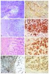
Renal cell carcinoma metastasizing to solitary fibrous tumor of the pleura: a case report
- PMID: 21714864
- PMCID: PMC3150316
- DOI: 10.1186/1752-1947-5-248
Renal cell carcinoma metastasizing to solitary fibrous tumor of the pleura: a case report
Abstract
Introduction: A tumor metastasizing to another malignancy is an uncommon phenomenon. Since it was first described in 1902, there have been fewer than 200 cases reported in the literature, with lung cancer metastasizing to renal cell carcinoma being the most frequently described pattern. Here we report a case of a solitary fibrous tumor of the lung acting as the recipient for a renal cell carcinoma. To our knowledge, this is the first reported case of such a combination and the second case involving a solitary fibrous tumor.
Case presentation: A 58-year-old Caucasian man who developed a persistent dry cough presented to our hospital. Imaging studies revealed a large pleural-based mass in the left lung. A biopsy of the mass showed a spindle-cell lesion consistent with a solitary fibrous tumor. The patient underwent surgical excision of the 13 cm mass. The pathological examination confirmed the diagnosis of a solitary fibrous tumor but also demonstrated discrete foci of metastatic renal cell carcinoma. Until that point, a primary renal cell carcinoma tissue diagnosis had not been made and the initial radiological work-up was inconclusive.
Conclusion: Awareness of the unusual phenomenon of tumor-to-tumor metastasis is important for practicing surgical pathologists, particularly in the evaluation of a mass lesion showing bimodal histology. This case also highlights the importance of careful examination of surgical specimens, as minute and unusual findings can direct patient care.
Figures


Similar articles
-
Renal malignant solitary fibrous tumor with single lymph node involvement: report of unusual metastasis and review of the literature.Onco Targets Ther. 2014 May 8;7:679-85. doi: 10.2147/OTT.S51664. eCollection 2014. Onco Targets Ther. 2014. PMID: 24855378 Free PMC article.
-
Small cell lung cancer associated with solitary fibrous tumors of the pleura: a case study and literature review.Int J Surg. 2014;12 Suppl 1:S19-21. doi: 10.1016/j.ijsu.2014.05.032. Epub 2014 May 22. Int J Surg. 2014. PMID: 24859397 Review.
-
Metachronous Malignant Solitary Fibrous Tumor of Kidney: Case Report and Review of Literature.Urol Case Rep. 2015 Oct 17;4:45-7. doi: 10.1016/j.eucr.2015.09.004. eCollection 2016 Jan. Urol Case Rep. 2015. PMID: 26793578 Free PMC article.
-
Folliculotropic Cutaneous Metastases and Lymphangitis Carcinomatosa: When Cutaneous Metastases of Breast Carcinoma Are Mistaken for Cutaneous Infections.Acta Dermatovenerol Croat. 2016 Jun;24(2):154-7. Acta Dermatovenerol Croat. 2016. PMID: 27477179
-
A giant solitary fibrous tumor of the pleura: diagnostic implications in an unusual case with literature review.Indian J Pathol Microbiol. 2010 Jul-Sep;53(3):544-7. doi: 10.4103/0377-4929.68289. Indian J Pathol Microbiol. 2010. PMID: 20699522 Review.
Cited by
-
Lung adenocarcinoma metastasizing to fibrous histiocytoma: A case report.Medicine (Baltimore). 2019 Jun;98(25):e16102. doi: 10.1097/MD.0000000000016102. Medicine (Baltimore). 2019. PMID: 31232953 Free PMC article.
-
Prevalence and risk factors of persistent cough in patients diagnosed with renal cell carcinoma: a systematic review and meta-analysis.BMJ Open. 2025 Mar 6;15(3):e088963. doi: 10.1136/bmjopen-2024-088963. BMJ Open. 2025. PMID: 40050050 Free PMC article.
-
Tumour-to-tumour metastasis: male breast carcinoma metastasis arising in an extrapleural solitary fibrous tumour - a case report.Diagn Pathol. 2014 Nov 25;9:203. doi: 10.1186/s13000-014-0203-y. Diagn Pathol. 2014. PMID: 25420931 Free PMC article.
-
Solitary pleural metastasis from renal cell carcinoma: a case of successful resection.Surg Case Rep. 2015;1(1):36. doi: 10.1186/s40792-015-0039-z. Epub 2015 Apr 23. Surg Case Rep. 2015. PMID: 26366340 Free PMC article.
-
Tumor-to-tumor metastasis: an unusual case of breast cancer metastatic to a solitary fibrous tumor.J Thorac Dis. 2016 Jun;8(6):E374-8. doi: 10.21037/jtd.2016.03.79. J Thorac Dis. 2016. PMID: 27293861 Free PMC article.
References
-
- Berent W. Seltene metastasenbildung. Zentralbl Allg Pathol. 1902;13:5.
LinkOut - more resources
Full Text Sources

