. 2011 Jun 29:8:75.
doi: 10.1186/1742-2094-8-75.

Lack of macrophage migration inhibitory factor in mice does not affect hallmarks of the inflammatory/immune response during the first week after stroke

Affiliations

Lack of macrophage migration inhibitory factor in mice does not affect hallmarks of the inflammatory/immune response during the first week after stroke

Ana R Inácio et al. J Neuroinflammation. .

Abstract

Background: Macrophage migration inhibitory factor (MIF) has been proposed to play a detrimental role in stroke. We recently showed that MIF promotes neuronal death and aggravates neurological deficits during the first week after experimental stroke, in mice. Since MIF regulates tissue inflammation, we studied the putative role of MIF in post-stroke inflammation.

Methods: We subjected C57BL/6 mice, Mif-/- (MIF-KO) or Mif+/+ (WT), to a transient occlusion of the right middle cerebral artery (tMCAo) or sham-surgery. We studied MIF expression, GFAP expression and the number of CD74-positive cells in the ischemic brain hemisphere 7 days after tMCAo using primarily immunohistochemistry. We determined IFN-γ, IL-2, IL-4, IL-5, IL-10, IL-12, KC/CXCL-1 and TNF-α protein levels in the brain (48 h after surgery) and serum (48 h and 7 days after surgery) by a multiplex immunoassay.

Results: We observed that MIF accumulates in neurons and astrocytes of the peri-infarct region, as well as in microglia/macrophages of the infarct core up to 7 days after stroke. Among the inflammatory mediators analyzed, we found a significant increase in cerebral IL-12 and KC levels after tMCAo, in comparison to sham-surgery. Importantly, the deletion of Mif did not significantly affect the levels of the cytokines evaluated, in the brain or serum. Moreover, the spleen weight 48 h and 7 days subsequent to tMCAo was similar in WT and MIF-KO mice. Finally, the extent of GFAP immunoreactivity and the number of MIF receptor (CD74)-positive cells within the ischemic brain hemisphere did not differ significantly between WT and MIF-KO mice subjected to tMCAo.

Conclusions: We conclude that MIF does not affect major components of the inflammatory/immune response during the first week after experimental stroke. Based on present and previous evidence, we propose that the deleterious MIF-mediated effects in stroke depend primarily on an intraneuronal and/or interneuronal action.


Figures

Figure 1
Figure 1
Experimental settings. (A) Composite photograph of a TTC stained brain 24 h after tMCAo. Representation of the cerebral damage obtained in average after a 45 min occlusion of the right middle cerebral artery, in C57BL/6 mice (white). Scale bar: 3 mm. (B) Experimental outline. IHC, immunohistochemistry.
Figure 2
Figure 2
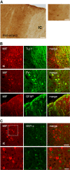
MIF accumulates in neurons and astrocytes of the peri-infarct region during the first 7 days after tMCAo, in C57BL/6 mice. (A) Bright field micrographs. Representative image of MIF immunoreactivity (MIF-ir) in the peri-infarct motor/somatosensory cortex at 7 days after tMCAo. Scale bar: 100 μm (right). A higher magnification image denotes the expression in neurons and glia-like cells. Note the thick glia-like processes positive for MIF around the infarct core. Scale bar: 50 μm (left). (B) Confocal micrographs. Co-localization of MIF (Cy3, red) with the neuronal markers Tuj-1 and PV (both Cy5, green). Co-localization of MIF (Cy3, red) and the marker GFAP (Cy5, green) confirms the presence in reactive astrocytes. Scale bar: 50 μm. Infarct core-IC. (C) Confocal micrographs. Lack of co-localization of MIF (Cy3, red) and GST-π (Cy5, green) in the peri-infarct somatosensory-motor cortex at 7 days after occlusion. Scale bars: 50 μm and 20 μm (lower and higher magnification, respectively).
Figure 3
Figure 3
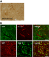
Expression of MIF in the infarct core of C57BL/6 mice at day 7 after tMCAo. (A) Representative bright field image of the striatal infarct core. Scale bar: 50 μm. (B) MIF is expressed in glia-like cells surrounding CD31+ cells, upper panel. Scale bar: 50 μm. Co-localization of MIF (Cy3, red) with the microglial/macrophage protein galectin-3(Gal-3)/Mac-2 (Alexa488, green), lower panel. Scale bar: 50 μm.
Figure 4
Figure 4
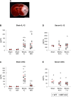
Genetic deletion of MIF does not affect the cerebral up-regulation of IL-12 and mKC 48 h after tMCAo, in mice. A) Schematic representation of the regions dissected from brains collected 48 h after surgery (sham and tMCAo): infarct core (IC, white) and non-infarct region (within dotted line). Scale bar: 3mm. B and D) IL-12 and mKC protein levels in the cerebral IC and non-infarct region (NIR) of WT and MIF-KO subjected to sham-operation or tMCAo. D and E) IL-12 and mKC protein levels in the serum of WT and MIF-KO 48 h and 7 days after sham-operation or tMCAo. In B, C, D and E we present individual data points and the mean (dash). # p < 0.05 tMCAo IC versus Sham IC (for each genotype). § p < 0.05 tMCAo NIR versus Sham NIR (for each genotype).
Figure 5
Figure 5
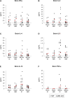
IFN-β, IL-2, IL-4, IL-5, IL-10 and TNF-α protein levels in the brains of WT and MIF-KO mice 48 h after tMCAo. Protein concentration in pg/mL; we present individual data points (as indicated in the figure) and the mean (dash). IC, infarct core; NIR, non-infarct region.
Figure 6
Figure 6
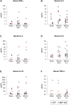
Serum IFN-β, IL-2, IL-4, IL-5, IL-10 and TNF-α protein levels 48 h and 7 days after tMCAo, in WT and MIF-KO mice. Protein concentration in pg/mL; we show individual data points (as indicated in the figure) and the mean (dash). IC, infarct core; P, peri-infarct region.
Figure 7
Figure 7
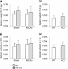
Spleen weight of WT and MIF-KO mice up to 7 days after tMCAo. Spleen weight (in g; mean ± STD) of WT and MIF-KO mice 48 h after either sham-surgery or tMCAo (A) and 7 days after tMCAo (A 1), and respective spleen weight (g)/body weight (g) ratios (B and B 1).
Figure 8
Figure 8
Cerebral GFAP immunoreactivity at day 7 post-tMCAo, in mice. Composite image representing the obtained GFAP immunoreactivity (Cy3, gray-scale) within the right brain hemisphere 7 days after a 45 min occlusion of the right middle cerebral artery, in C57BL/6 mice (epifluorescence micrographs).
Figure 9
Figure 9
Number of CD74+ cells in WT and MIF-KO brains 7 days after tMCAo. (A and A1) Epifluorescence micrographs. (A) Representative composite image of CD74 immunoreactivity in the mouse brain 7 days post-occlusion (WT). Note the presence of CD74+ cells essentially within the infarct core. (A1) Higher magnification micrographs denote the morphology of CD74+ cells. Scale bar: 10 μm. (B) Confocal images. Lack of co-localization MIF (Alexa 488, green)/CD74 (Cy3, red) in the infarct core. Scale bars: 50 μm and 10 μm (lower and higher magnification, respectively). (C) Number of CD74+ cells detected per ipsilateral brain hemisphere (sum of the analyzed brain levels) - WT and MIF-KO mice 7 days post-occlusion of the MCA (mean ± STD).

Similar articles

Cited by

References

    1. Endres M, Engelhardt B, Koistinaho J, Lindvall O, Meairs S, Mohr JP, Planas A, Rothwell N, Schwaninger M, Schwab ME, Vivien D, Wieloch T, Dirnagl U. Improving outcome after stroke: overcoming the translational roadblock. Cerebrovasc Dis. 2008;25:268–78. doi: 10.1159/000118039. - DOI - PubMed
    1. Wang Q, Tang XN, Yenari MA. The inflammatory response in stroke. J Neuroimmunol. 2007;184:53–68. doi: 10.1016/j.jneuroim.2006.11.014. - DOI - PMC - PubMed
    1. Denes A, Thornton P, Rothwell NJ, Allan SM. Inflammation and brain injury: acute cerebral ischaemia, peripheral and central inflammation. Brain Behav Immun. 2010;24:708–23. doi: 10.1016/j.bbi.2009.09.010. - DOI - PubMed
    1. Liu T, McDonnell PC, Young PR, White RF, Siren AL, Hallenbeck JM, Barone FC, Feurestein GZ. Interleukin-1 beta mRNA expression in ischemic rat cortex. Stroke. 1993;24:1746–50. - PubMed
    1. Yamasaki Y, Matsuura N, Shozuhara H, Onodera H, Itoyama Y, Kogure K. Interleukin-1 as a pathogenetic mediator of ischemic brain damage in rats. Stroke. 1995;26:676–80. - PubMed

Publication types

MeSH terms