Review
. 2011 Sep 22;118(12):3212-21.
doi: 10.1182/blood-2011-02-306597. Epub 2011 Jun 29.

Unraveling the scissile bond: how ADAMTS13 recognizes and cleaves von Willebrand factor

Affiliations
Review

Unraveling the scissile bond: how ADAMTS13 recognizes and cleaves von Willebrand factor

James T B Crawley et al. Blood. .

Abstract

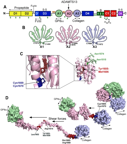
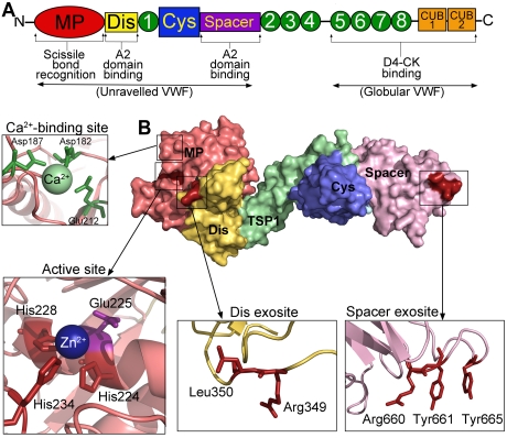
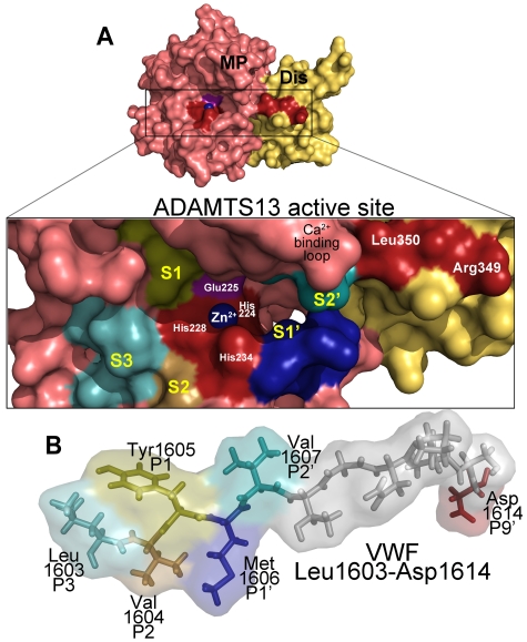
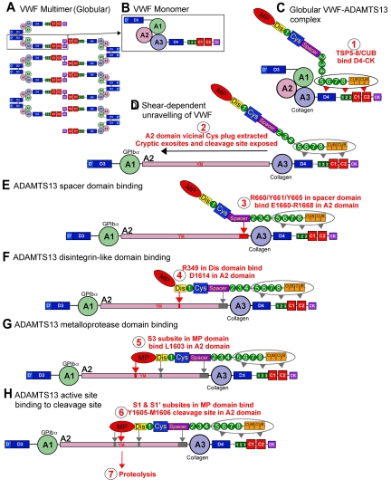
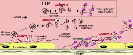
von Willebrand factor (VWF) is a large adhesive glycoprotein with established functions in hemostasis. It serves as a carrier for factor VIII and acts as a vascular damage sensor by attracting platelets to sites of vessel injury. VWF size is important for this latter function, with larger multimers being more hemostatically active. Functional imbalance in multimer size can variously cause microvascular thrombosis or bleeding. The regulation of VWF multimeric size and platelet-tethering function is carried out by ADAMTS13, a plasma metalloprotease that is constitutively active. Unusually, protease activity of ADAMTS13 is controlled not by natural inhibitors but by conformational changes in its substrate, which are induced when VWF is subject to elevated rheologic shear forces. This transforms VWF from a globular to an elongated protein. This conformational transformation unfolds the VWF A2 domain and reveals cryptic exosites as well as the scissile bond. To enable VWF proteolysis, ADAMTS13 makes multiple interactions that bring the protease to the substrate and position it to engage with the cleavage site as this becomes exposed by shear. This article reviews recent literature on the interaction between these 2 multidomain proteins and provides a summary model to explain proteolytic regulation of VWF by ADAMTS13.


Figures

Figure 1
Figure 1
The substrate, VWF. (A) Domain organization of VWF. N-linked and O-linked glycosylation sites are represented by green and orange lollipops, respectively. The propeptide (yellow represents D1 and D2) is marked. S-S indicates sites of intermolecular disulphide bond pairing. Vertical arrows indicate the location of cleavage sites (furin and ADAMTS13). Ligand binding sites (FVIII, GPIbα, collagen, and αIIbβIII) are labeled below. (B) Schematic representation of the VWF A1, A2, and A3 domains. The paired cysteines in A1 and A3 are shown. The vicinal disulphide bond that forms the molecular plug in the A2 domain and the Tyr1605-Met1606 (YM) ADAMTS13 cleavage site are also represented. (C) Structure of the VWF A2 domain highlighting the N-linked glycosylation sites (green), vicinal disulphide bond (blue), and the ADAMTS13 cleavage site (red) hidden in the center of the folded domain. (D) Molecular models of the unfolding of the VWF A1-A2-A3 domains. In globular VWF, the A3 domain collagen binding site is exposed. Elevated shear forces on VWF cause uncoupling of the A domains, extraction of the Cys1669-Cys1670 vicinal disulphide plug, and unraveling of the A2 domain. This exposes the GPIbα binding site in the A1 domain, cryptic ADAMTS13 binding sites, and the cleavage site in the A2 domain (red).
Figure 2
Figure 2
The protease, ADAMTS13. (A) Domain organization of ADAMTS13. From the N-terminus are the metalloprotease domain (MP; red), disintegrin-like domain (Dis; yellow), TSP repeats (1-8; green), cysteine-rich domain (Cys; blue), spacer domain (purple), and CUB domains (orange). Binding sites and function of specific domains are labeled below. (B) Structure of ADAMTS13 N-terminal domains (MDTCS) based on the crystal structure of DTCS and homology modeling of the MP domain. Surface representation is shown. Domains are colored according to panel A. (Insets) Cartoon representation of the location of the high-affinity calcium binding site and coordinating residues (green) in the MP domain, the active site containing 3 His residues (red) and catalytic Glu225 (mauve), the disintegrin-like domain exosite, and spacer domain exosite.
Figure 3
Figure 3
ADAMTS13 recognition of the VWF cleavage site. (A) Molecular model of the ADAMTS13 metalloprotease (MP; light red) and disintegrin-like (Dis; yellow) domains. (Inset) The active site cleft. Active site residues (Zn2+ ion, its 3 coordinating His residues His224, His228, and His234, as well as the catalytic Glu225 residue) and disintegrin-like domain exosite residues are labeled. The high-affinity functional Ca2+-binding loop is also marked. Regions of the metalloprotease domain that are predicted to harbor the S3, S2, S1, S1′, and S2′ subsites are labeled marked in different colors. (B) Below the active site cleft, VWF A2 domain residues Leu1603-Asp1614 are depicted as sticks and transparent spacefill. The P3, P2, P1, P1′, P2′, and P9′ residues in VWF that are important for proteolysis are labeled and colored according to their predicted complementary subsites (S) in the metalloprotease domain.
Figure 4
Figure 4
Proteolysis of VWF by ADAMTS13. VWF circulates in plasma as a multimeric molecule (A) that adopts a quiescent globular conformation. Each multimer is composed of disulphide linked VWF monomers (B). In its globular conformation, the A3 domain collagen binding site is exposed. ADAMTS13 can bind to this globular VWF via its TSP (5-8) and CUB domains (C), step 1. This enables VWF and ADAMTS13 complexes to form and circulate in plasma. Under elevated shear forces (which can occur on secretion, collagen binding, or passage through the microvasculature), VWF can unravel to expose A1 domain binding site for GPIbα. These shear forces also remove molecular plug formed by the vicinal disulphide bond in the A2 domain, which causes A2 domain unfolding (D), step 2 (see Figure 1). This unfolding reveals cryptic exosites that enable residues in the ADAMTS13 spacer domain to bind to the unfolded A2 domain (E), step 3 (see Figures 1D, 2B). Thereafter, a critical low-affinity interaction between D1614 and the Dis domain helps approximate and position the cleavage site (F), step 4 (see Figure 3). This enables further interactions between the MP domain to occur, including an essential interaction via an S3 subsite with L1603 in VWF (G), step 5 (see Figure 3). Together, these interactions allow the MP to engage via S1 and S1′ subsites with the cleavage site (YM; H), step 6, after which proteolysis can occur, step 7.
Figure 5
Figure 5
Location of VWF cleavage by ADAMTS13. Cartoon depicting the sites of VWF proteolysis by ADAMTS13. (UL)VWF is synthesized by the endothelium and stored within Weibel-Palade bodies (WPB; green). VWF multimers of various sizes, including UL-VWF, can be secreted directly into the circulation. (1) Alternatively, a proportion of UL-VWF may attach to the endothelial surface during exocytosis and unravel in response to shear forces. Under such circumstances, the VWF A2 domain unfolds to enable ADAMTS13 (red scissors) to cleave VWF and release the VWF string. Whether directly secreted or proteolytically released, VWF can adopt a globular fold in the plasma circulation. However, during passage through the microvasculature, globular UL-VWF in free circulation will probably unravel (at least partially/transiently). (2) Such unraveling permits the processing of the largest, most hemostatically active forms of VWF, resulting in their conversion to smaller plasma VWF multimers. Mutations in VWF that precipitate type 2A von Willebrand disease are particularly influenced by such proteolysis. This group of mutations enhances the propensity of VWF to unfold in free circulation, leading to excessive proteolysis and loss of hemostatically functional VWF. Conversely, ADAMTS13 deficiency results in the loss of such plasma processing. Under these circumstances, platelets (Pl) can become bound to transiently unraveled VWF, leading to the accumulation of VWF-platelet aggregates that occlude the microvasculature, as seen in patients presenting with TTP. At sites of vessel damage, endothelial damage results in exposure of subendothelial collagen. Plasma VWF binds to this, unravels, and, in turn, recruits platelets. The presence of collagen and thrombin induces rapid platelet activation, which consolidates the platelet plug. Thrombin further stabilizes this through the deposition of fibrin and the proteolytic inactivation of ADAMTS13. (3) Downstream of the site of injury (ie, in the absence of collagen and thrombin), VWF-platelet strings may still be proteolysed by ADAMTS13, which in turn limits/regulates platelet plug formation. Low or reduced ADAMTS13 levels may impair this process and consequently influence the pathophysiology of arterial thrombosis.

References

    1. Sadler JE. Biochemistry and genetics of von Willebrand factor. Annu Rev Biochem. 1998;67:395–424. - PubMed
    1. Ruggeri ZM, De Marco L, Gatti L, Bader R, Montgomery RR. Platelets have more than one binding site for von Willebrand factor. J Clin Invest. 1983;72(1):1–12. - PMC - PubMed
    1. Bockenstedt P, Greenberg JM, Handin RI. Structural basis of von Willebrand factor binding to platelet glycoprotein Ib and collagen: effects of disulfide reduction and limited proteolysis of polymeric von Willebrand factor. J Clin Invest. 1986;77(3):743–749. - PMC - PubMed
    1. Roth GJ, Titani K, Hoyer LW, Hickey MJ. Localization of binding sites within human von Willebrand factor for monomeric type III collagen. Biochemistry. 1986;25(26):8357–8361. - PubMed
    1. Hamer RJ, Koedam JA, Beeser-Visser NH, Bertina RM, Van Mourik JA, Sixma JJ. Factor VIII binds to von Willebrand factor via its Mr-80,000 light chain. Eur J Biochem. 1987;166(1):37–43. - PubMed

Publication types

MeSH terms