. 2011 Sep 1;39(17):7791-802.
doi: 10.1093/nar/gkr509. Epub 2011 Jun 28.

Translation-competent 48S complex formation on HCV IRES requires the RNA-binding protein NSAP1

Affiliations

Translation-competent 48S complex formation on HCV IRES requires the RNA-binding protein NSAP1

Sung Mi Park et al. Nucleic Acids Res. .

Abstract

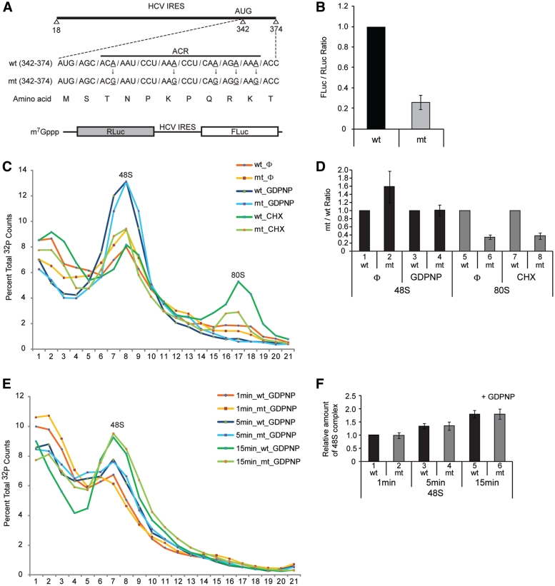
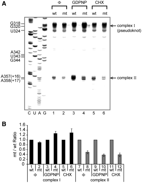
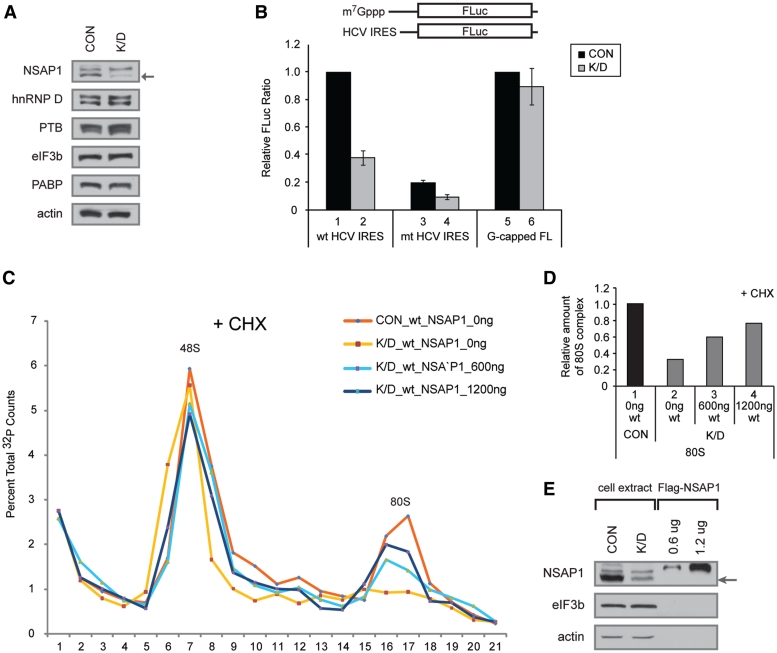
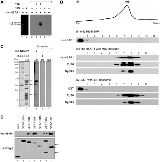
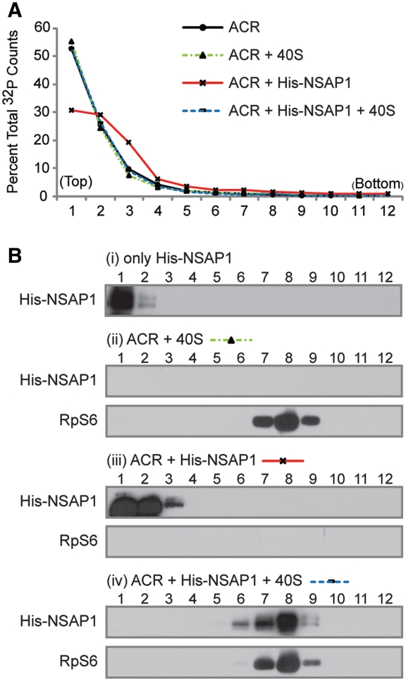
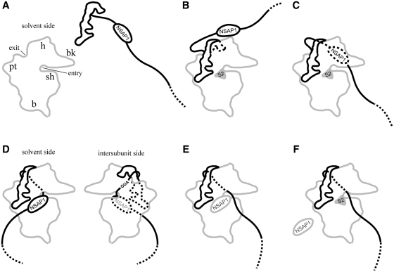
Translation of many cellular and viral mRNAs is directed by internal ribosomal entry sites (IRESs). Several proteins that enhance IRES activity through interactions with IRES elements have been discovered. However, the molecular basis for the IRES-activating function of the IRES-binding proteins remains unknown. Here, we report that NS1-associated protein 1 (NSAP1), which augments several cellular and viral IRES activities, enhances hepatitis C viral (HCV) IRES function by facilitating the formation of translation-competent 48S ribosome-mRNA complex. NSAP1, which is associated with the solvent side of the 40S ribosomal subunit, enhances 80S complex formation through correct positioning of HCV mRNA on the 40S ribosomal subunit. NSAP1 seems to accomplish this positioning function by directly binding to both a specific site in the mRNA downstream of the initiation codon and a 40S ribosomal protein (or proteins).


Figures

Figure 1.
Figure 1.
NSAP1 facilitates 80S complex formation. (A) Schematic diagram of the NSAP1-binding site on the HCV IRES. wt and mt, which contains A–G substitutions that impede NSAP1-binding of HCV IRES RNAs, are depicted. (B) Translational efficiencies of wt and mt HCV IRESs were measured in RRLs using dicistronic mRNAs (lower panel in A). The relative HCV IRES translational efficiencies are depicted. (C) Representative sucrose gradient data of three experiments. The translation reaction mixtures were incubated in RRLs at 30°C for 15 min with [32P]-labeled wt or mt HCV IRES RNAs (18–374 nt) in the presence or absence of GDPNP or CHX. Pre-initiation complex and initiation complex on the HCV IRESs were observed using 5–20% sucrose gradient analyses. Φ denotes no drug treatment. (D) Relative amount of 48S and 80S complexes on wt or mt HCV IRES quantified from panel C. The radioactivity of each complex (48S or 80S complex) was quantified by using manual planimetry after subtraction of the basal radioactivity from the radioactivity of each fraction. The mean and standard deviation values of three independent experiments are depicted by columns and bars. The complexes on wt HCV IRES were assigned a value of 1 (lanes 1, 3, 5 and 7). (E) Representative sucrose gradient data of three experiments. The translation reaction mixtures were incubated in RRLs at 30°C for 1, 5 and 15 min with [32P]-labeled wt or mt HCV IRES RNAs (18–374 nt) in the presence of GDPNP. The 48S complex on the HCV IRESs was observed using 5–20% sucrose gradient analyses. (F) Relative amount of 48S complex on wt or mt HCV IRES was measured from panel E similarly to panel D. The amount of 48S complex formed on wt HCV IRES in 1 min was assigned a value of 1.
Figure 2.
Figure 2.
NSAP1 promotes the correct positioning of the 40S ribosomal subunit at the initiation codon. (A) Toeprinting analyses of wt and mt HCV IRESs in RRLs. Representative toeprint data of five independent experiments. The toeprints of wt and mt HCV RNAs in the absence of RRL are presented in Supplementary Figure S2B, which shows that the differences between toeprints are not due to mutations in the HCV RNA per se. (B) Relative amount of complexes I and II on wt or mt HCV IRES quantified from five experiments. The band intensities on the gel shown in panel A were quantified by autoradiography, and the relative amounts of the complexes on mt IRES to those on wt IRES at various experimental settings are depicted. The complexes on wt HCV IRES were assigned a value of 1 (lanes 1, 3, 5, 7, 9 and 11). The mean and standard deviation values of five independent experiments are depicted by columns and bars.
Figure 3.
Figure 3.
Depletion of NSAP1 inhibits 80S complex formation on the HCV IRES. (A) Western blot analyses of lysates from control 293T cells (CON) and 293T cells treated with an siRNA against NSAP1 (K/D). (B) Relative FLuc activity directed from monocistronic reporter RNAs bearing wt HCV IRES or m7G-capped reporter (G-capped FL) in 293T cell lysates. The luciferase activity from wt HCV IRES in control cells (lane 1 for lanes 1–4) and that from capped RNA in control cells (lane 5 for lanes 5 and 6) were assigned a value of 1. (C) The effect of NSAP1 on 80S complex formation was monitored by sucrose gradient analyses of NSAP1-depleted and NSAP1-replete lysates. The labeled RNAs were incubated in control (CON) or NSAP1-depleted lysates (K/D) in the presence of CHX, and then subjected to sucrose gradient analyses. The effect of NSAP1 supplementation on 80S complex formation was observed by adding purified Flag-NSAP1 protein (600 or 1200 ng) to NSAP1-depleted lysates. The radioactivity [32P-labeled wt IRES (18–374 nt)] in each fraction of the 5–20% sucrose gradient is depicted as a percentile in the graph. (D) Relative amount of 80S complex on wt HCV IRES quantified from panel C. (E) The amount of NSAP1 in control (NSAP1-replete) lysates, NSAP1-depleted lysates and NSAP1-depleted lysates supplemented with purified NSAP1 was monitored by western blotting.
Figure 4.
Figure 4.
NSAP1 directly interacts with the purified 40S ribosomal subunit in vitro. (A) Co-migration of 40S ribosomal subunit and NSAP1 on an agarose gel. Purified His-NSAP1 proteins were incubated with purified 40S or 60S ribosomal subunits and resolved on a 1% agarose gel. The positions of ribosomes and NSAP protein were observed by ethidium bromide staining (lanes 1 and 2) or western blotting with an antibody against the His-tag (lanes 3–7). (B) Co-migration of the 40S ribosomal subunit and His-NSAP1 in 5–20% sucrose density gradients. After centrifugation, the absorbance of the gradient at 254 nm was monitored using a Bio-Rad EM-1 UV monitor (panel i), and the gradient was fractionated into 11 samples. The presence of proteins in each fraction was monitored by western blotting (panels ii–iv). (C) Detection of NSAP1-interacting r-proteins by far-western blotting. (D) Confirmation of protein–protein interactions by GST-pull down assay using purified GST-fused r-proteins and His-NSAP1. Arrows depict proteins derived from GST-RpS2 and GST-RpS6 in protein purification process. RpS2, RpS4, RpS6 and RpS8 were shown to interact with NSAP1.
Figure 5.
Figure 5.
The fate of NSAP1 on the HCV IRES after interacting with the 40S ribosomal subunit. (A) Radioactivity profile of ACR (NSAP1-binding site). ACR: ACR RNA alone; ACR + 40S: ACR RNA incubated with purified 40S ribosomal subunits for 10 min; ACR + NSAP1: ACR RNA incubated with purified NSAP1 for 10 min; ACR + NSAP1 + 40S: ACR RNA incubated with His-NSAP1 for 10 min, followed by addition of purified 40S ribosomal subunits to the mixture. The reaction mixtures were subjected to sucrose gradient analyses. (B) Western blot analyses of proteins in each fraction shown in (A).
Figure 6.
Figure 6.
Proposed model for the enhancement of HCV mRNA translation by NSAP1. (A) NSAP1 binds to ACR on the HCV IRES. (B) HCV IRES associates with the 40S ribosomal subunit. (C) The coding region of HCV mRNA is in motion to find the P-site in the 40S ribosomal subunit. (D) NSAP1 interacts with a r-protein (or proteins) on the solvent side of the 40S ribosomal subunit. This is predicted to result in positioning of the AUG initiation codon at the P-site of the ribosome. (E) NSAP1 is released from the ACR and translation commences. (F) NSAP1 is released from the 40S ribosomal subunit, becoming available for use in the next round of translation. (B, C and D–F) represent complexes I and II in Figure 2, respectively.

References

    1. Jackson RJ, Hellen CU, Pestova TV. The mechanism of eukaryotic translation initiation and principles of its regulation. Nat. Rev. Mol. Cell. Biol. 2010;11:113–127. - PMC - PubMed
    1. Pelletier J, Sonenberg N. Internal initiation of translation of eukaryotic mRNA directed by a sequence derived from poliovirus RNA. Nature. 1988;334:320–325. - PubMed
    1. Jang SK, Krausslich HG, Nicklin MJ, Duke GM, Palmenberg AC, Wimmer E. A segment of the 5′ nontranslated region of encephalomyocarditis virus RNA directs internal entry of ribosomes during in vitro translation. J. Virol. 1988;62:2636–2643. - PMC - PubMed
    1. Sonenberg N, Hinnebusch AG. Regulation of translation initiation in eukaryotes: mechanisms and biological targets. Cell. 2009;136:731–745. - PMC - PubMed
    1. Pestova TV, Hellen CU, Shatsky IN. Canonical eukaryotic initiation factors determine initiation of translation by internal ribosomal entry. Mol. Cell. Biol. 1996;16:6859–6869. - PMC - PubMed

Publication types

MeSH terms