. 2011 Sep;85(17):8841-51.
doi: 10.1128/JVI.00539-11. Epub 2011 Jun 29.

Adenovirus E1A directly targets the E2F/DP-1 complex

Affiliations

Adenovirus E1A directly targets the E2F/DP-1 complex

Peter Pelka et al. J Virol. 2011 Sep.

Abstract

Deregulation of the cell cycle is of paramount importance during adenovirus infection. Adenovirus normally infects quiescent cells and must initiate the cell cycle in order to propagate itself. The pRb family of proteins controls entry into the cell cycle by interacting with and repressing transcriptional activation by the E2F transcription factors. The viral E1A proteins indirectly activate E2F-dependent transcription and cell cycle entry, in part, by interacting with pRb and family members to free the E2Fs. We report here that an E1A 13S isoform can unexpectedly activate E2F-responsive gene expression independently of binding to the pRb family of proteins. We demonstrate that E1A binds to E2F/DP-1 complexes through a direct interaction with DP-1. E1A appears to utilize this binding to recruit itself to E2F-regulated promoters, and this allows the E1A 13S protein, but not the E1A 12S protein, to activate transcription independently of interaction with pRb. Importantly, expression of E1A 13S, but not E1A 12S, led to significant enhancement of E2F4 occupancy of E2F sites of two E2F-regulated promoters. These observations identify a novel mechanism by which adenovirus deregulates the cell cycle and suggest that E1A 13S may selectively activate a subset of E2F-regulated cellular genes during infection.


Figures

Fig. 1.
Fig. 1.
E1A 13S induces pRb-independent activation of E2F-mediated transcription and binds E2F/DP-1 complexes. (A) U2OS cells were cotransfected with the reporter plasmid pGL-E2F (having four synthetic consensus E2F binding sites) together with E2F1 and DP-1 and the indicated E1A constructs or the vector alone. Luciferase assays were performed 48 h after transfection, and results were normalized to the protein concentration. Data are plotted as the n-fold increase over the reporter alone. (B) A U2OS cell line harboring an integrated (“chromatinized”) pGL-E2F reporter was cotransfected with the indicated E2Fs together with DP-1 and either E1A 12S dl1108 or E1A 13S dl1108. Luciferase activity was measured 48 h after transfection and is represented as n-fold activation versus that in cells transfected only with empty plasmids (vector = 1). (C) HeLa cells were transfected with plasmids expressing HA-tagged E2F1 to E2F5, HA-tagged DP-1, and E1A 13S dl1108 as indicated. Forty-eight hours after transfection, cells were lysed and immunoprecipitations for E1A were carried out using M73 anti-E1A monoclonal antibody. Immunoprecipitates were resolved on SDS-PAGE, and associated E2F/DP-1 complexes were detected by Western blotting using anti-HA antibody (3F10). Inputs of E2Fs, DP-1, and E1A are shown.
Fig. 2.
Fig. 2.
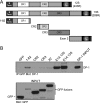
E1A N terminus binds to DP-1. (A) Schematic representation of E1A-GFP fusion fragments used in the experiment. (B) HeLa cells were cotransfected with the indicated GFP-E1A fragment fusions or with E1A 12S or E1A 13S together with HA-tagged DP-1. Either GFP or E1A (in the cases of E1A 12S and E1A 13S) was immunoprecipitated (IP) from cell lysates and resolved by SDS-PAGE, and associated DP-1 was detected using anti-HA antibody (3F10). Inputs of DP-1 and GFP fusion proteins are shown.
Fig. 3.
Fig. 3.
E1A residues 2 to 11 are required for pRb-independent association with DP-1. (A) Schematic representation of E1A exon 1 showing the mutant forms used in the study. N-Term., N terminus. (B) HeLa cells were cotransfected with vectors expressing the indicated E1A 12S deletion variants and HA–DP-1. Cells were lysed 48 h after transfection, and immunoprecipitations (IP) were carried out using anti-E1A antibody (M73). Associated DP-1 was detected using anti-HA antibody (3F10). Input levels are indicated. (C) HeLa cells were cotransfected with vectors expressing the indicated E1A proteins and HA–DP-1. Cells were lysed 48 h after transfection, and E1A immunoprecipitations were carried out using anti-E1A antibody (M73). Associated DP-1 was detected using anti-HA antibody (3F10). Input levels are indicated. (D) HeLa cells were cotransfected with vectors expressing the indicated E1A proteins and HA–DP-1. Cells were lysed 48 h after transfection, and E1A was immunoprecipitated with anti-E1A antibody (M73). Associated DP-1 was detected using anti-HA antibody (3F10). Input levels are shown. wt, wild type.
Fig. 4.
Fig. 4.
DP-1 N terminus is required for association with E1A. (A) Schematic representation of DP-1 and deletion mutant forms used in this study. (B, left) HeLa cells were cotransfected with either E1A 12S or E1A 13S and the indicated deletion mutant forms of DP-1. Cells were lysed 48 h after transfection, and E1A was immunoprecipitated (IP) using anti-E1A antibody (M73). Associated DP-1 was detected using anti-HA antibody (3F10). (B, right) HeLa cells were cotransfected with the indicated deletion mutant forms of DP-1 and the Y47H/C124G double mutant form of E1A 12S that is unable to bind to pRb. Cells were lysed 48 h after transfection, and E1A was immunoprecipitated using anti-E1A antibody (M73). Associated DP-1 was detected using anti-HA antibody (3F10). Inputs are shown for all immunoprecipitations. wt, wild type.
Fig. 5.
Fig. 5.
E1A binds endogenous DP-1 during viral infection. (A) IMR-90 cells were infected with adenoviruses expressing the indicated E1A isoforms (no E1A [Mock], E1A 12S, E1A 12S Δ2-11 Y47H/C124G, or E1A 13S). Twenty-four hours after infection, cells were lysed and E1A was immunoprecipitated (IP) using anti-E1A (M73) antibody. Associated DP-1 was detected using anti-DP-1 antibody (WTH24). Each experiment was done twice from two independent infections. Inputs of DP-1 and E1As are shown. (B) IMR-90 cells were infected with adenoviruses expressing the indicated E1A isoforms (no E1A [Mock], wild-type genomic E1A [dl309], wild-type E1A 13S, or E1A 13S Y47H/C124G Δ2-11). Twenty-four hours after infection, the cells were lysed and E1A was immunoprecipitated using anti-E1A (M73) antibody. Associated endogenous E2F4 was detected using a C-terminal E2F4 antibody. Inputs of E2F4 and E1A are shown.
Fig. 6.
Fig. 6.
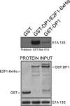
E1A 13S binds directly to DP-1/E2F1 and DP-1 alone. GST pulldown assays were carried out with the purified DP-1/E2F1 complex and purified E1A 13S or DP-1 and E1A 13S alone. GST was used as a negative control. E1A pulled down by DP-1/E2F1 or DP-1 alone was detected using anti-E1A (M73) antibody. Inputs are shown.
Fig. 7.
Fig. 7.
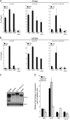
E1A is recruited to E2F-regulated promoters, altering E2F4 occupancy and E2F target gene expression. (A) ChIPs and re-ChIPs were carried out for E1A (using M73 antibody), E2F4 C-terminal antibody, or IgG control antibody (mouse anti-rabbit) in arrested IMR-90 cells infected with adenovirus with E1 deleted, virus expressing only E1A 12S (dl520), virus expressing only E1A 13S (pm975), or virus expressing genomic E1A (both E1A 12S and 13S; dl309). Immunoprecipitations were carried out 24 h after infection. Occupancy of E1A and E2F4 was assessed for the PCNA promoter around E2F sites by qRT-PCR and is expressed as a percentage of the input. (B) ChIPs and re-ChIPs were carried out for E1A (using M58 and M73 antibodies), E2F4 (using a custom rabbit E2F4 antibody), or IgG control antibody (mouse anti-rabbit) in arrested IMR-90 cells infected with either adenovirus with E1 deleted, virus expressing only E1A 12S (dl520), virus expressing only E1A 13S (pm975), or virus expressing genomic E1A (both E1A 12S and 13S; dl309). Immunoprecipitations were carried out 24 h after infection. Occupancy of E1A and E2F4 was assessed for the MCM4 promoter around E2F sites using qRT-PCR and is expressed as a percentage of the input. (C) Expression levels of various E1A mutant adenoviruses in infected IMR-90 cells 24 h after infection. Arrested IMR-90 cells were infected with the indicated viruses and lysed, and E1A levels were detected using anti-E1A (M73) antibody. Actin was used as a loading control. (D) Real-time PCR quantification of expression levels of the PCNA, MCM4, and BLM gene products at 36 h after infection of arrested IMR-90 cells. The results are expressed as n-fold changes versus cells infected with adenovirus with E1 deleted.
Fig. 8.
Fig. 8.
CR3 and the N-terminal DP-1-binding domain are required for efficient induction of S phase and virus growth. (A) Arrested IMR-90 cells were infected with the indicated adenoviruses, pulsed with BrdU for 1 h at the indicated time points, fixed, and stained with anti-BrdU antibody. Results are expressed as a percentage of BrdU-positive nuclei and constitute a mean of three random fields of view at a low magnification. (B) Arrested IMR-90 cells were infected with pm975 or dl520 virus at an MOI of 5, and medium was assayed for cell-free virus 48 and 120 h after infection by a plaque assay on 293 cells.

References

    1. Avvakumov N., Kajon A. E., Hoeben R. C., Mymryk J. S. 2004. Comprehensive sequence analysis of the E1A proteins of human and simian adenoviruses. Virology 329:477–492 - PubMed
    1. Bayley S. T., Mymryk J. S. 1994. Adenovirus E1A proteins and transformation. Int. J. Oncol. 5:425–444 - PubMed
    1. Berk A. J. 2005. Recent lessons in gene expression, cell cycle control, and cell biology from adenovirus. Oncogene 24:7673–7685 - PubMed
    1. Blais A., Dynlacht B. D. 2004. Hitting their targets: an emerging picture of E2F and cell cycle control. Curr. Opin. Genet. Dev. 14:527–532 - PubMed
    1. Bruton R. K., et al. 2008. Identification of a second CtBP binding site in adenovirus type 5 E1A conserved region 3. J. Virol. 82:8476–8486 - PMC - PubMed

Publication types

LinkOut - more resources