. 2011 Sep;85(17):8954-67.
doi: 10.1128/JVI.00754-11. Epub 2011 Jun 29.

Mechanism of neutralization by the broadly neutralizing HIV-1 monoclonal antibody VRC01

Affiliations

Mechanism of neutralization by the broadly neutralizing HIV-1 monoclonal antibody VRC01

Yuxing Li et al. J Virol. 2011 Sep.

Abstract

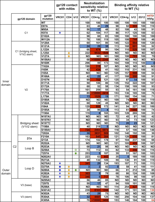
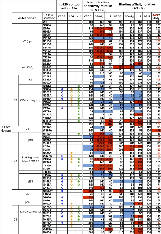
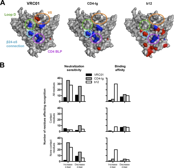
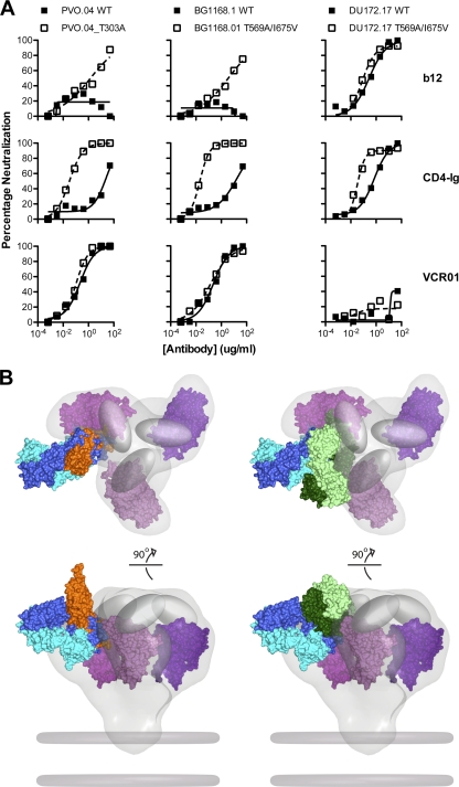
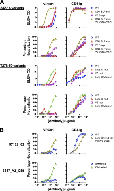
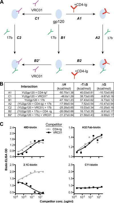
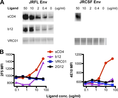
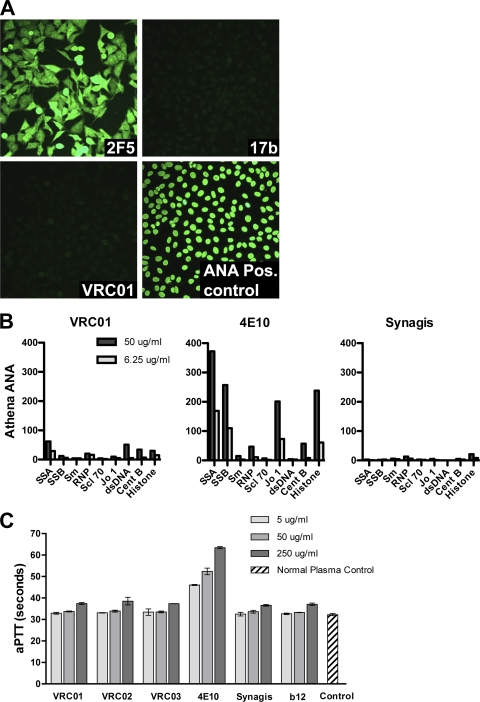
The structure of VRC01 in complex with the HIV-1 gp120 core reveals that this broadly neutralizing CD4 binding site (CD4bs) antibody partially mimics the interaction of the primary virus receptor, CD4, with gp120. Here, we extended the investigation of the VRC01-gp120 core interaction to the biologically relevant viral spike to better understand the mechanism of VRC01-mediated neutralization and to define viral elements associated with neutralization resistance. In contrast to the interaction of CD4 or the CD4bs monoclonal antibody (MAb) b12 with the HIV-1 envelope glycoprotein (Env), occlusion of the VRC01 epitope by quaternary constraints was not a major factor limiting neutralization. Mutagenesis studies indicated that VRC01 contacts within the gp120 loop D, the CD4 binding loop, and the V5 region were necessary for optimal VRC01 neutralization, as suggested by the crystal structure. In contrast to interactions with the soluble gp120 monomer, VRC01 interaction with the native viral spike did not occur in a CD4-like manner; VRC01 did not induce gp120 shedding from the Env spike or enhance gp41 membrane proximal external region (MPER)-directed antibody binding to the Env spike. Finally, VRC01 did not display significant reactivity with human antigens, boding well for potential in vivo applications. The data indicate that VRC01 interacts with gp120 in the context of the functional spike in a manner distinct from that of CD4. It achieves potent neutralization by precisely targeting the CD4bs without requiring alterations of Env spike configuration and by avoiding steric constraints imposed by the quaternary structure of the functional Env spike.


Figures

Fig. 1.
Fig. 1.
VRC01 neutralization and binding on a panel of primary isolate JRCSF functional Env and gp120 mutants. Amino acid numbering of mutants is based on the HIV-1 HXBc2 sequence. The gp120 structurally defined contact residues for the VRC01, CD4, and b12 complexes are indicated in blue, orange, and green, respectively, with open circles (○) designating gp120 main-chain-only contacts, asterisk (*) designating gp120 side chain-only contacts, and filled circles (●) designating both main-chain and side-chain contacts. Mutations that knock out putative N-glycosylation sites (NXS/T motifs) of gp120 are labeled with a dagger symbol (†). Relative ELISA binding affinities to captured gp120s were calculated based on the antibody concentration at half-maximal binding (EC50). The effect of each mutation on antibody binding was normalized using 2G12 to control for the amount of captured gp120. Mutations that resulted in decreased gp120 binding (<33% relative to that of wild type [WT]) are highlighted in blue, and those that resulted in increased ELISA binding (>300% relative to that for the wild type) are highlighted in red. HIVIg or the anti-V3 MAb 447D (values shown in red) were used as the controls for gp120s with poor 2G12 binding. Neutralization sensitivity of each mutant to MAb VRC01, CD4-Ig, or MAb b12 was assessed and compared to neutralization of the wild-type Env pseudovirus. The color scheme is the same as that used for the gp120 binding data.
Fig. 1.
Fig. 1.
VRC01 neutralization and binding on a panel of primary isolate JRCSF functional Env and gp120 mutants. Amino acid numbering of mutants is based on the HIV-1 HXBc2 sequence. The gp120 structurally defined contact residues for the VRC01, CD4, and b12 complexes are indicated in blue, orange, and green, respectively, with open circles (○) designating gp120 main-chain-only contacts, asterisk (*) designating gp120 side chain-only contacts, and filled circles (●) designating both main-chain and side-chain contacts. Mutations that knock out putative N-glycosylation sites (NXS/T motifs) of gp120 are labeled with a dagger symbol (†). Relative ELISA binding affinities to captured gp120s were calculated based on the antibody concentration at half-maximal binding (EC50). The effect of each mutation on antibody binding was normalized using 2G12 to control for the amount of captured gp120. Mutations that resulted in decreased gp120 binding (<33% relative to that of wild type [WT]) are highlighted in blue, and those that resulted in increased ELISA binding (>300% relative to that for the wild type) are highlighted in red. HIVIg or the anti-V3 MAb 447D (values shown in red) were used as the controls for gp120s with poor 2G12 binding. Neutralization sensitivity of each mutant to MAb VRC01, CD4-Ig, or MAb b12 was assessed and compared to neutralization of the wild-type Env pseudovirus. The color scheme is the same as that used for the gp120 binding data.
Fig. 2.
Fig. 2.
(A) Surface model of the JRCSF gp120 core showing mutants resulting in decreased binding (blue) or increased binding (red). The gp120 domains that contain mutations most affecting affinities for VRC01 are denoted in circles of the following colors: green, loop D; gold, V5/β24; purple, CD4 BLP (CD4 binding loop); and blue, β24-α5 connection. (B) Histograms showing the number of mutant residues affecting virus neutralization or gp120 binding for VRC01, b12, and CD4-Ig.
Fig. 3.
Fig. 3.
Impact of conformational constraints on neutralization by b12, CD4-Ig, and VRC01. (A) Env pseudoviruses PVO.04 (left) and BG1168.1 (middle) are relatively resistant to b12 and CD4-Ig. In the setting of the gp120 T303A mutation or the gp41 T569A/I675V mutations that result in a more open Env conformation, these viruses show enhanced neutralization sensitivity to b12 and CD4-Ig. In contrast, both the wild-type and mutant versions of these viruses were equally sensitive to VRC01-mediated neutralization. Virus DU172.17 (right) was resistant to VRC01, and this resistance was not affected by the Env mutations that alter conformation. (B) Model of VRC01, CD4, and b12 binding to the HIV-1 functional spike. Top (top) and front (bottom) views of the density map surface of the HIV-1 spike, presenting three gp120 core protomers in magenta (39) (Electron Microscopy Database [EMDB]: EMD-5019; Protein Data Bank [PDB]: 3DNN) and viral membrane in dark gray. To analyze the relative binding orientations of b12, CD4, and VRC01, the X-ray coordinates for gp120, in the b12, VRC01, and CD4/17b/bound conformations (PDB: 2NY7, 3NGB, and 2NY3), respectively, were superimposed on the same gp120 core protomer (PDB: 3DNN). The molecular surface of the b12-Fab fragment is green; the VRC01 Fab is blue. Within each color-coded Fab, the heavy chains are shaded darker than the light chains. The molecular surface of sCD4 is orange, and the variable loops are represented as gray ovals. Note that in this model, b12 and CD4 are more loop proximal than is VRC01.
Fig. 4.
Fig. 4.
Binding and neutralization of reversion mutants and chimeras of VRC01-resistant viruses. (A) VRC01-resistant viruses 242-14 and T278-50 and their reversion mutants. Curves show binding and neutralization with VRC01 or CD4-Ig. The specific reversion mutants and chimeras are described in Table 2. (B) Analysis of two Env pseudoviruses that were resistant to VRC01 despite recognition of the viral gp120. gp120 of virus 57128_02 bound to VRC01 with an EC50 of 8.7 μg/ml (Table 1) yet was resistant to VRC01 neutralization. With additional reversion mutations in loop D, CD4 binding loop, and V5, the mutant virus was moderately neutralized by VRC01 (top). Virus 3817_V2_C59 was resistant to VRC01 neutralization (bottom) despite strong recognition of its gp120 by VRC01 (EC50 of 0.04 μg/ml, Table 1). When the virus was produced in the presence of the glycan modification inhibitor kifunensin, it became sensitive to VRC01-mediated neutralization (bottom).
Fig. 5.
Fig. 5.
VRC01 effects on monomeric gp120, compared with CD4-Ig. (A) Scheme of thermodynamic cycles showing enhancement of binding to gp120 between CD4bs ligands CD4-Ig and VRC01 and the CD4i antibody 17b. (B) Thermodynamic values of CD4-Ig, VRC01, and 17b interactions with gp120 measured by ITC at 37°C, including the change in enthalpy (ΔH) and entropy (−TΔS) upon binding of gp120 to the coreceptor mimetic antibody 17b, antibody VRC01, and CD4-Ig, a surrogate for the primary receptor, CD4. *, data extracted from reference . (C) Effect on other CD4i antibodies binding to gp120, captured by sheep anti-gp120 C5 antibody D7324 on ELISA plate. The binding of biotin-labeled CD4i antibodies, 48D, A32 Fab, and 2.1C to gp120 was tested in the presence of CD4-Ig or VRC01, with signals detected by the streptavidin-HRP conjugate. The MAb C11, which recognizes a C1-to-C5 conformational epitope on gp120, was used as negative control.
Fig. 6.
Fig. 6.
VRC01 effects on HIV envelope glycoprotein functional trimer. (A) Western blot analysis of Env from supernatants of 293T cells transfected with cleavage competent gp160 JRFL or JRCSF Env plasmid DNA. HIV-1 gp120 shedding upon binding of ligands, including sCD4, b12, and VRC01, from the Env trimer on the surface of 293T cells was evaluated by probing the blot with the anti-gp120 human monoclonal antibody 39F (V3 specific). (B) Effect of VRC01, b12, and sCD4 on gp41 MPER-directed antibodies 2F5 and 4E10 binding to trimeric Env. 293T cells transfected with JRFL-ΔCT Env expression plasmid were stained with biotin-labeled 2F5 or 4E10. Binding of 2F5 or 4E10 to JRFL Env in the presence of the CD4bs ligands VRC01, b12, and sCD4 was detected with streptavidin-APC conjugate. MFI, mean fluorescence intensity.
Fig. 7.
Fig. 7.
Lack of VRC01 self-reactivity. (A) Reactivity of MAbs 2F5, 17b, and VRC01 with human HEp-2 epithelial cells. The MAb 2F5 served as a positive control and reacted in a diffuse cytoplasmic and nuclear pattern. (B) Luminex AtheNA Multi-Lyte ANA test for a panel of nuclear antigens: systemic lupus erythematosus autoantigens SSA and SSB, sphingomyelin (Sm), ribonucleoprotein (RNP), sclerosis autoantigen (Scl-70), histidine-tRNA ligase (Jo-1), double-stranded DNA (dsDNA), and centromere B (CentB). (C) The clinical assay, activated partial thromboplastin time (aPTT), was used to assess potential antiplatelet, antiphospholipid activity. The MAbs were added to normal sera, and the time to clot formation was measured. The clotting times for VRC01, VRC02, VRC03, b12, and the anti-RSV monoclonal antibody Synagis (palivizumab) were all in the normal range. In contrast, the known polyreactive MAb 4E10 caused a prolongation of the aPTT.

References

    1. Alkhatib G., et al. 1996. CC CKR5: a RANTES, MIP-1alpha, MIP-1beta receptor as a fusion cofactor for macrophage-tropic HIV-1. Science 272:1955–1958 - PubMed
    1. Binley J. M., et al. 2010. Role of complex carbohydrates in human immunodeficiency virus type 1 infection and resistance to antibody neutralization. J. Virol. 84:5637–5655 - PMC - PubMed
    1. Binley J. M., et al. 2008. Profiling the specificity of neutralizing antibodies in a large panel of plasmas from patients chronically infected with human immunodeficiency virus type 1 subtypes B and C. J. Virol. 82:11651–11668 - PMC - PubMed
    1. Blish C. A., Nguyen M. A., Overbaugh J. 2008. Enhancing exposure of HIV-1 neutralization epitopes through mutations in gp41. PLoS Med. 5:e9. - PMC - PubMed
    1. Buchacher A., et al. 1994. Generation of human monoclonal antibodies against HIV-1 proteins; electrofusion and Epstein-Barr virus transformation for peripheral blood lymphocyte immortalization. AIDS Res. Hum. Retroviruses 10:359–369 - PubMed

Publication types

MeSH terms

Substances