. 2011 Sep;85(17):9147-58.
doi: 10.1128/JVI.00121-11. Epub 2011 Jun 29.

Passive immunotherapies protect WRvFire and IHD-J-Luc vaccinia virus-infected mice from lethality by reducing viral loads in the upper respiratory tract and internal organs

Affiliations

Passive immunotherapies protect WRvFire and IHD-J-Luc vaccinia virus-infected mice from lethality by reducing viral loads in the upper respiratory tract and internal organs

Marina Zaitseva et al. J Virol. 2011 Sep.

Abstract

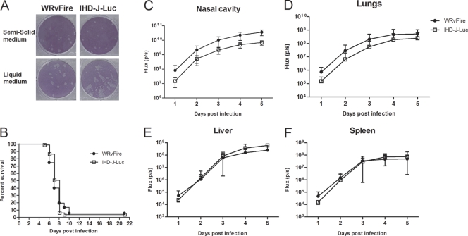
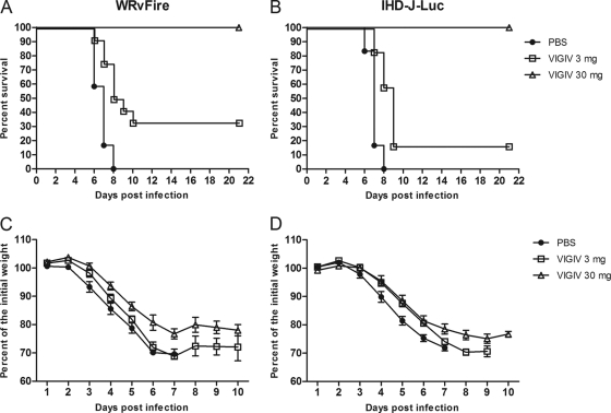
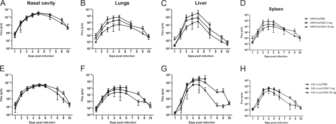
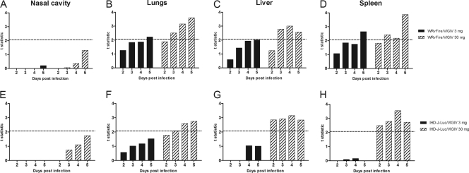
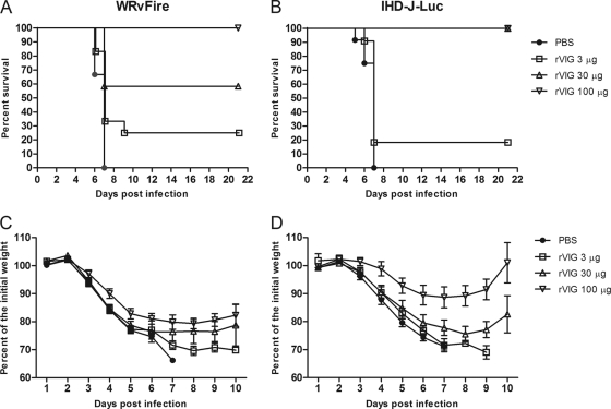
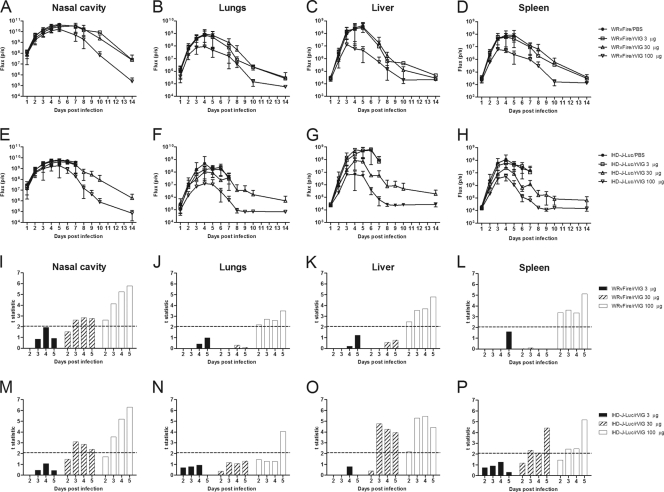
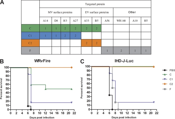
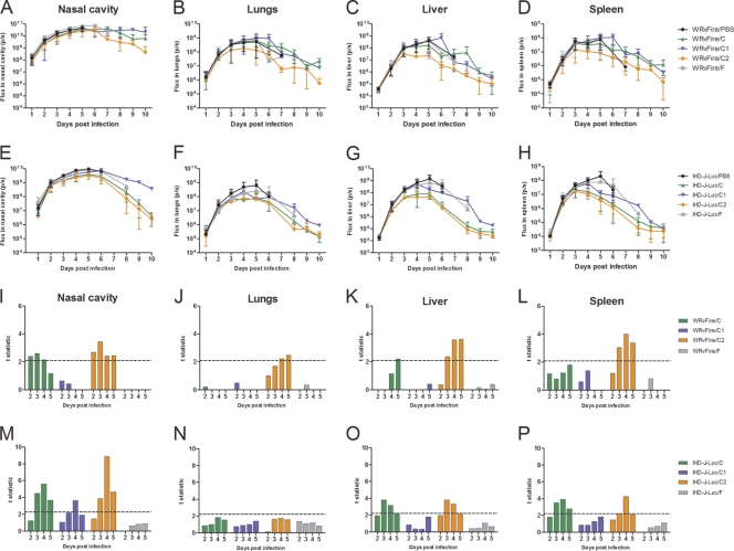
Whole-body bioimaging was employed to study the effects of passive immunotherapies on lethality and viral dissemination in BALB/c mice challenged with recombinant vaccinia viruses expressing luciferase. WRvFire and IHD-J-Luc vaccinia viruses induced lethality with similar times to death following intranasal infection, but WRvFire replicated at higher levels than IHD-J-Luc in the upper and lower respiratory tracts. Three types of therapies were tested: licensed human anti-vaccinia virus immunoglobulin intravenous (VIGIV); recombinant anti-vaccinia virus immunoglobulin (rVIG; Symphogen, Denmark), an investigational product containing a mixture of 26 human monoclonal antibodies (HuMAbs) against mature virion (MV) and enveloped virion (EV); and HuMAb compositions targeting subsets of MV or EV proteins. Bioluminescence recorded daily showed that pretreatment with VIGIV (30 mg) or with rVIG (100 μg) on day -2 protected mice from death but did not prevent viral replication at the site of inoculation and dissemination to internal organs. Compositions containing HuMAbs against MV or EV proteins were protective in both infection models at 100 μg per animal, but at 30 μg, only anti-EV antibodies conferred protection. Importantly, the t statistic of the mean total fluxes revealed that viral loads in surviving mice were significantly reduced in at least 3 sites for 3 consecutive days (days 3 to 5) postchallenge, while significant reduction for 1 or 2 days in any individual site did not confer protection. Our data suggest that reduction of viral replication at multiple sites, including respiratory tract, spleen, and liver, as monitored by whole-body bioluminescence can be used to predict the effectiveness of passive immunotherapies in mouse models.


Figures

Fig. 1.
Fig. 1.
In vitro infection of mouse cells and infection of mice in vivo with WRvFire and IHD-J-Luc vaccinia viruses. (A) Visualization of plaques formed by WRvFire and IHD-J-Luc viruses. Monolayers of 3T3 mouse cells were infected with WrvFire (left panels) or with IHD-J-Luc (right panels), overlaid with solid (top panels) or liquid (bottom panels) medium, and stained with crystal violet. (B to F) Lethality outcome and bioluminescence measurements of recombinant WRvFire and IHD-J-Luc vaccinia viruses in the internal organs of BALB/c mice. Eighty-seven and 81 BALB/c mice were infected i.n. with WRvFire (circles) and with IHD-J-Luc (squares) vaccinia viruses, respectively. Mice were observed for survival (B) and were subjected to whole-body bioimaging (C to F). Total fluxes in the nasal cavity (C), lungs (D), liver (E), and spleen (F) were determined as described in materials and methods and used to calculate mean total flux ± SD using t test.
Fig. 2.
Fig. 2.
VIGIV protected mice from lethality following WRvFire or IHD-J-Luc infections in a dose-dependent manner. Mice were inoculated i.p. with VIGIV at 3 mg/animal (squares), 30 mg/animal (triangles), or PBS (closed circles) 2 days before i.n. challenge with WRvFire (A and C) and with IHD-J-Luc (B and D) vaccinia viruses. Mice were observed for survival (A and B) for 21 days postinfection; weight measurements were performed daily for 10 days and used to calculate mean percentages of initial weight ± SD (C and D). For the VIGIV (30 mg)-treated and IHDJ-Luc-infected groups, n = 11; for all other groups, n = 12. Results are representative of three experiments.
Fig. 3.
Fig. 3.
Bioluminescence in organs of mice pretreated with VIGIV prior to infections with WRvFire or IHD-J-Luc. Mice were inoculated i.p. with VIGIV (day −2) at 3 mg/animal (squares), 30 mg/animal (triangles), or with PBS (closed circles), and were infected i.n. (day 0) with WRvFire (A-D) or IHD-J-Luc vaccinia viruses (E-H). Animals were subjected to whole-body imaging for 10 days. Bioluminescence in the nasal cavity (A, E), lungs (B, F), liver (C, G), and spleen (D, H) was recorded and used to calculate mean total flux ± SD. For the numbers of animals per groups see legend to Fig. 2.
Fig. 4.
Fig. 4.
Statistical analysis of total fluxes recorded in organs of WRvFire- or IHD-J-Luc-infected control mice and mice treated with VIGIV. Total fluxes were recorded in the nasal cavity (A and E), lungs (B and F), liver (C and G), and spleen (D and H) of mice infected with WRvFire (A to D) or with IHD-J-Luc (E to H). The y axis shows t values that depict differences between the means of the total fluxes in control mice (PBS treated) and the means of the total fluxes in animals that received 3 mg/animal (closed bars) or 30 mg/animal (hatched bars) of VIGIV. Dashed horizontal lines depict the critical t values of 2.07 (12 mice per group) (A to D) and 2.08 (11 or 12 mice per group) (E to H). All values on or above the dashed lines are significant at α = 0.05 (two tailed).
Fig. 5.
Fig. 5.
rVIG protects mice from lethal challenge with WRvFire and IHD-J-Luc in a dose-dependent manner. BALB/c mice were inoculated with rVIG at 3 μg/animal (squares), 30 μg/animal (triangles), or 100 μg/animal (inverted triangles) or with PBS (closed circles) and 2 days later challenged with WRvFire (A) or IHD-J-Luc (B). Mice were observed for lethality for 21 days; weight measurements were performed daily for 10 days and used to calculate mean percentages of initial weight ± SD (C and D). For the PBS-treated and WRvFire-challenged groups, n = 15. For all rVIG-treated groups infected with WRvFire, n = 12. For the PBS-treated and for rVIG (30 μg)-treated and IHDJ-Luc-challenged groups, n = 12. For the 3-μg and 100-μg rVIG-treated IHDJ-Luc-infected groups, n = 11. Results are representative of three experiments.
Fig. 6.
Fig. 6.
Bioluminescence in mice pretreated with rVIG prior to infections with WRvFire or IHD-J-Luc and statistical analysis of total fluxes in organs. (A to H) Bioluminescence of infected mice. Mice were inoculated i.p. with rVIG at 3 μg/animal (squares), 30 μg/animal (triangles), or 100 μg/animal (inverted triangles) or with PBS (closed circles) 2 days before i.n. challenge with WRvFire (A to D) or IHDJ-Luc (E to H). Animals were subjected to whole-body imaging for 10 days to record bioluminescence and calculate mean total flux ± SD in the nasal cavity (A and E), lungs (B and F), liver (C and G), and spleen (D and H). (I to P) Statistical analysis. Mean total fluxes were used to calculate the t statistic between groups of mice treated with 3 μg/animal (solid bars), 30 μg/animal (hatched bars), or 100 μg/animal (open bars) of rVIG and PBS. The dashed horizontal line depicts the critical t values of 2.06 in the WRvFire challenge (12 to 15 mice per group) (I to L) and 2.08 in the IHD-J-Luc challenge (11 or 12 mice per group) (M to P).
Fig. 7.
Fig. 7.
Effects of individual HuMAb compositions on lethality in WRvFire challenge model. (A) Specificities of HuMAbs targeting MV, EV surface vaccinia virus proteins and nonsurface (other) proteins within individual compositions are shown in the table. Numbers of MAbs against a specific target are indicated in colored cells. (B and C) BALB/c mice were inoculated i.p. with HuMAb compositions C (green triangles), C1 (blue inverted triangles), C2 (orange diamonds), and F (gray squares) at 30 μg/animal or with PBS (black circles) and were challenged with WRvFire (B) or with IHD-J-Luc (C) viruses 2 days later. Six to 12 mice per group were used in the experiment. Mice were observed for lethality and survival for 21 days. Results are representative of three experiments.
Fig. 8.
Fig. 8.
Bioluminescence and statistical analysis of total fluxes in mice that received prophylactic treatment with HuMAb compositions and were challenged with WRvFire or with IHD-J-Luc vaccinia virus. (A to H) Bioluminescence of infected mice. BALB/c mice were inoculated i.p. with HuMAb compositions C (green triangles), C1 (blue inverted triangles), C2 (orange diamonds), and F (gray squares) at 30 μg/animal or with PBS (black circles) and were challenged with WRvFire (A-D) or with IHD-J-Luc (E-H) viruses 2 days later. Animals were subjected to whole-body imaging for 10 days to record bioluminescence and calculate mean total flux ± SD in the nasal cavity (A and E), lungs (B and F), liver (C and G), and spleen (D and H). (I to P) Statistical analysis. Mean total fluxes were used to calculate the t statistic between groups of mice treated with compositions C (green bars), C1 (blue bars), C2 (orange bars), and F (gray bars) and PBS-treated mice. The dashed horizontal lines depict critical t values of 2.09 in the WRvFire challenge (10 to 12 mice per group) (I to L) and 2.28 in the IHD-J-Luc challenge (6 mice per group) (M to P).

References

    1. Bell E., et al. 2004. Antibodies against the extracellular enveloped virus B5R protein are mainly responsible for the EEV neutralizing capacity of vaccinia immune globulin. Virology 325:425–431 - PubMed
    1. Benhnia M. R., et al. 2009. Vaccinia virus extracellular enveloped virion neutralization in vitro and protection in vivo depend on complement. J. Virol. 83:1201–1215 - PMC - PubMed
    1. Benhnia M. R., et al. 2008. Redundancy and plasticity of neutralizing antibody responses are cornerstone attributes of the human immune response to the smallpox vaccine. J. Virol. 82:3751–3768 - PMC - PubMed
    1. Blasco R., Moss B. 1992. Role of cell-associated enveloped vaccinia virus in cell-to-cell spread. J. Virol. 66:4170–4179 - PMC - PubMed
    1. Cono J., Casey C. G., Bell D. M. 2003. Smallpox vaccination and adverse reactions. Guidance for clinicians. MMWR Recomm. Rep. 52:1–28 - PubMed

Publication types

MeSH terms

LinkOut - more resources