. 2011 Jun 29;31(26):9456-65.
doi: 10.1523/JNEUROSCI.1460-11.2011.

Human immunodeficiency virus infection of human astrocytes disrupts blood-brain barrier integrity by a gap junction-dependent mechanism

Affiliations

Human immunodeficiency virus infection of human astrocytes disrupts blood-brain barrier integrity by a gap junction-dependent mechanism

Eliseo A Eugenin et al. J Neurosci. .

Abstract

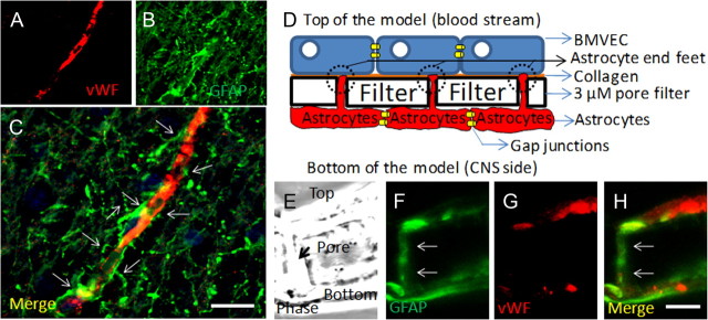
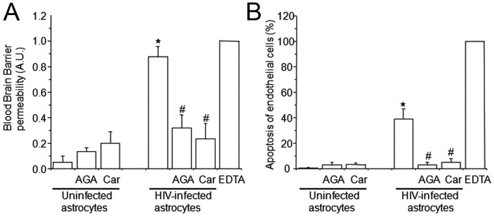
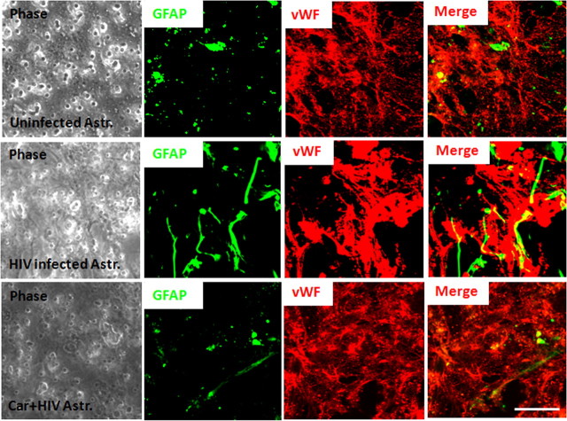
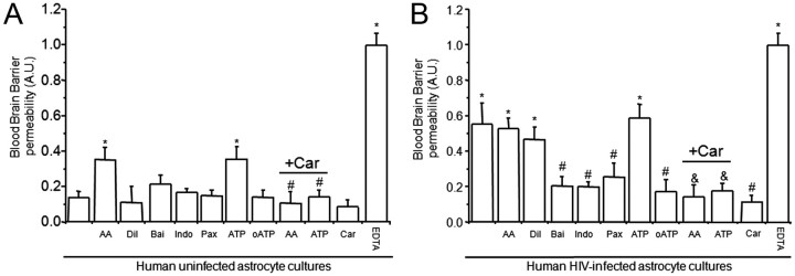
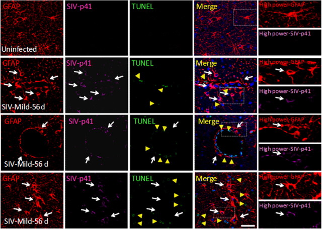
HIV infection of the CNS is an early event after primary infection, resulting in neurological complications in a significant number of individuals despite antiretroviral therapy (ART). The main cells infected with HIV within the CNS are macrophages/microglia and a small fraction of astrocytes. The role of these few infected astrocytes in the pathogenesis of neuroAIDS has not been examined extensively. Here, we demonstrate that few HIV-infected astrocytes (4.7 ± 2.8% in vitro and 8.2 ± 3.9% in vivo) compromise blood-brain barrier (BBB) integrity. This BBB disruption is due to endothelial apoptosis, misguided astrocyte end feet, and dysregulation of lipoxygenase/cyclooxygenase, BK(Ca) channels, and ATP receptor activation within astrocytes. All of these alterations in BBB integrity induced by a few HIV-infected astrocytes were gap junction dependent, as blocking these channels protected the BBB from HIV-infected astrocyte-mediated compromise. We also demonstrated apoptosis in vivo of BBB cells in contact with infected astrocytes using brain tissue sections from simian immunodeficiency virus-infected macaques as a model of neuroAIDS, suggesting an important role for these few infected astrocytes in the CNS damage seen with HIV infection. Our findings describe a novel mechanism of bystander BBB toxicity mediated by low numbers of HIV-infected astrocytes and amplified by gap junctions. This mechanism of toxicity contributes to understanding how CNS damage is spread even in the current ART era and how minimal or controlled HIV infection still results in cognitive impairment in a large population of infected individuals.


Figures

Figure 1.
Figure 1.
Astrocytes are key cells in the maintenance of the BBB in vivo and in vitro. A–C, Staining of human brain tissue sections for vWF (a marker of endothelial cells; red staining) (A) and GFAP (a marker of astrocytes; green staining) (B) demonstrates the close interaction between astrocytes and their end feet with endothelial cells in vivo (C) (merge of both colors). D, Schematic of our in vitro model of the human BBB, composed of BMVECs grown on denatured collagen on the top of a polycarbonate membrane with 3 μm pores, and human astrocytes cultured on the bottom side of the membrane. Astrocytes that are coupled by gap junctions send processes, termed astrocyte end feet, through the 3 μm pores. E–H, Staining of a cross section of the BBB model (see phase picture; E) for GFAP (F) or vWF (G) or the merge of both colors demonstrated that vWF staining is in the top of the insert and GFAP-positive astrocytes are on the bottom. Astrocytes send processes from the bottom of the insert to the top of the insert to establish contact with the BMVEC layer. Much of this GFAP staining is also detectable on the top of the insert (H; colocalization of both colors; yellow). Thus, our in vitro model of the human BBB recapitulates many of the astrocyte–endothelial interactions seen in vivo. This model will be used to examine the role of HIV-infected astrocytes in BBB integrity and astrocyte end feet signaling. Scale bar, 50 μm.
Figure 2.
Figure 2.
The use of HIV-infected astrocyte cultures to establish the human BBB model results in BBB permeability (A) and endothelial apoptosis (B). BBB disruption was evaluated by the permeability of the model to albumin conjugated to Evans blue dye, and endothelial apoptosis was evaluated by TUNEL. A, BBB models using uninfected astrocytes were totally impermeable to albumin conjugated to Evans blue dye. Gap junction blockers AGA or Car did not alter BBB integrity when uninfected astrocytes cultures were used to establish the model. The BBB established using HIVADA-infected astrocyte cultures had disrupted integrity, despite no detectable infection of astrocyte cultures as assayed by p24 ELISA. Addition of gap junction blockers AGA (32 μm) or Car (10 μm) in the bottom chamber on the BBB model (astrocyte side) inhibited BBB disruption. EDTA added to the BBB model was used as a positive control for disruption (n = 4; *p ≤ 0.005 compared with control conditions using uninfected astrocytes; #p ≤ 0.005 compared with the BBB model using HIV-infected astrocytes). B, Determination of endothelial apoptosis using TUNEL staining and confocal microscopy of control BBB cultures indicated no apoptosis. Gap junction blockers AGA or Car, used in treating control BBB cultures established with uninfected astrocytes, did not cause endothelial apoptosis. BBB models with HIVADA-infected astrocyte cultures exhibit significantly increased endothelial apoptosis. Addition of GJ blockers AGA or Car (10 μm) in the bottom chamber of the BBB model (astrocyte side) totally abolished endothelial apoptosis induced by few HIV-infected astrocytes (n = 4; *p ≤ 0.005 compared with control conditions; #p ≤ 0.005 compared with BBB using HIV-infected astrocytes).
Figure 3.
Figure 3.
Astrocyte end feet are dysregulated in the presence of HIV-infected astrocytes resulting in BBB disruption and permeability. Confocal microscopy was performed on the endothelial layer (vWF and EC marker; red staining) to observe the astrocyte end feet (GFAP, an astrocyte marker; green staining) in contact with the monolayer of endothelial cells. In control conditions, localized spots of astrocyte end feet were detected in contact with the monolayer of BMVECs. The use of HIV-infected astrocyte cultures in the BBB model resulted in random localization and aberrant formation of astrocyte end feet in contact with a disrupted BMVEC. The addition of the GJ blocker, carbenoxolone (10 μm), to the bottom of the inserts (astrocyte side), resulted in mostly normal appearing astrocyte end feet processes in contact with the monolayer of BMVECs and protection against disruption of the BMVEC monolayer (n = 3). Phase of the inserts shows the pores of the filters. Scale bar, 20 μm.
Figure 4.
Figure 4.
Astrocyte end feet signaling participate in BBB disruption mediated by few HIV-infected astrocytes. We examined whether normal signaling pathways concentrated in astrocyte end feet that regulate blood flow are altered by HIVADA infection of astrocytes. We determined whether activation or blocking pathways used to control blood flow in the brain altered BBB integrity of our tissue culture model using albumin conjugated to Evans blue dye to assay for permeability. A, BBB cultures established with BMVECs and uninfected human astrocytes were totally impermeable. The addition of AA or ATP to the bottom of the BBB cultures established using uninfected astrocytes induced partial BBB disruption. Blocking activation of lipoxygenase and cyclooxygenase with baicalein (Bai) and indomethacin (Indo) on the astrocyte side did not alter BBB permeability when uninfected astrocytes were using. Blocking conductance calcium-activated potassium channels (BKCa) with paxilline (Pax) in the bottom of the BBB model, astrocyte side, did not alter permeability. Blocking ATP purinergic receptors using oxidized ATP (oATP) did not alter permeability of the barrier when uninfected astrocytes were used for the barrier. The disruption of the BBB induced by AA and ATP was GJ dependent because carbenoxolone (+Car) abolished disruption (n = 4; *p ≤ 0.005 compared with control and #p ≤ 0.003 compared with BBB inserts treated with AA or ATP). B, BBB cultures established using BMVECs and HIV-infected human astrocytes were highly permeable. The addition of AA, the diluent of AA (Tocrisolve), or ATP to the bottom of the BBB cultures did not change the already disrupted BBB. Blocking activation of lipoxygenase and cyclooxygenase using Bai and Indo on the astrocyte side was protective against BBB disruption induced by few infected astrocytes. Blocking BKCa with Pax in the bottom of the BBB model was also protective. Blocking ATP purinergic receptors using oATP (10 μm) was protective. This suggests the involvement of several signaling pathways in the BBB disruption induced by few HIV-infected astrocytes. The addition of carbenoxolone (+Car) to the bottom of the BBB cultures abolished BBB disruption induced by AA, ATP, or HIV-infected astrocytes (as shown in Fig. 2), indicating that gap junction channels play a key role in amplifying astrocyte end feet dysregulation resulting in BMVEC compromise and permeability (n = 4; *p < 0.005 compared with control uninfected conditions; #p < 0.003 compared with permeable BBB cultures established using HIV-infected astrocyte cultures; &p < 0.005 compared with treatment of BBB cultures containing HIV-infected astrocytes, treated with AA or ATP).
Figure 5.
Figure 5.
SIV-infected astrocytes are detected in vivo (8.2 ± 3.9%) and are found in close proximity to apoptotic neighboring cells. SIV-infected astrocytes are not apoptotic. Confocal analyses and subsequent three-dimensional reconstructions were performed using tissue sections obtained from uninfected and mildly encephalitic macaques 21–84 d after SIV infection. Tissue was stained for nuclei (DAPI; blue), GFAP (astrocyte marker; red), TUNEL (apoptosis; green), and SIV-p41 (SIV; magenta). Uninfected tissue sections showed uniform astrocyte staining with extensive processes and no apoptosis and no staining for SIV-p41 protein. Staining of brain tissue sections obtained from 21 and 56 d after infection showed 8.2 ± 3.9% infected astrocytes. Animals with mild encephalitis at 56 d after infection had significant infection of astrocytes (white arrows) and apoptosis in neighboring uninfected endothelial cells (yellow arrows, second row) and astrocytes (yellow arrows, third row). SIV-infected astrocytes did not apoptose. The white box in the merge pictures represents the area amplified in the last column to demonstrate colocalization of GFAP and SIV-p41 staining (arrows indicated SIV-infected astrocytes). In severe encephalitis (data not shown) due to the significant loss of parenchymal organization and extensive inflammation, it was not possible to assay reliably for SIV infection of astrocytes or apoptosis in neighboring cells. Scale bar, 40 μm.

Similar articles

Cited by

References

    1. Abbott NJ. Astrocyte-endothelial interactions and blood-brain barrier permeability. J Anat. 2002;200:629–638. - PMC - PubMed
    1. Acheampong EA, Parveen Z, Muthoga LW, Kalayeh M, Mukhtar M, Pomerantz RJ. Human immunodeficiency virus type 1 Nef potently induces apoptosis in primary human brain microvascular endothelial cells via the activation of caspases. J Virol. 2005;79:4257–4269. - PMC - PubMed
    1. Anthony IC, Ramage SN, Carnie FW, Simmonds P, Bell JE. Influence of HAART on HIV-related CNS disease and neuroinflammation. J Neuropathol Exp Neurol. 2005;64:529–536. - PubMed
    1. Babas T, Vieler E, Hauer DA, Adams RJ, Tarwater PM, Fox K, Clements JE, Zink MC. Pathogenesis of SIV pneumonia: selective replication of viral genotypes in the lung. Virology. 2001;287:371–381. - PubMed
    1. Bagasra O, Lavi E, Bobroski L, Khalili K, Pestaner JP, Tawadros R, Pomerantz RJ. Cellular reservoirs of HIV-1 in the central nervous system of infected individuals: identification by the combination of in situ polymerase chain reaction and immunohistochemistry. AIDS. 1996;10:573–585. - PubMed

Publication types

MeSH terms